Разделы презентаций
- Разное
- Английский язык
- Астрономия
- Алгебра
- Биология
- География
- Геометрия
- Детские презентации
- Информатика
- История
- Литература
- Математика
- Медицина
- Менеджмент
- Музыка
- МХК
- Немецкий язык
- ОБЖ
- Обществознание
- Окружающий мир
- Педагогика
- Русский язык
- Технология
- Физика
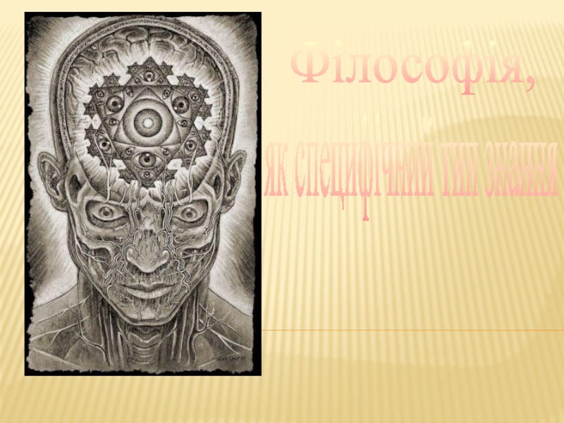
- Философия
- Химия
- Шаблоны, картинки для презентаций
- Экология
- Экономика
- Юриспруденция
Anatomy of the human brain
Содержание
- 1. Anatomy of the human brain
- 2. Anatomy of the human brainThe Lateral Surface
- 3. Anatomy of the human brainCerebral Lobes and
- 4. Anatomy of the human brainSelected Gyri, Sulci,
- 5. Anatomy of the human brainSelected Gyri, Sulci,
- 6. Anatomy of the human brainCerebral Lobes and
- 7. Anatomy of the human brainMajor Sensory, Motor,
- 8. Anatomy of the human brainMajor Sensory, Motor,
- 9. Anatomy of the human brainMajor Sensory, Motor,
- 10. Anatomy of the human brainMajor Sensory, Motor,
- 11. Слайд 11
- 12. Anatomy of the human brainThe Medial Surface
- 13. Anatomy of the human brainForebrain StructuresCorpus callosum
- 14. Anatomy of the human brainForebrain StructuresThe amygdala
- 15. Anatomy of the human brainVentriclesthe third ventriclethe cerebral aqueductthe fourth ventriclethe spinal canal
- 16. Anatomy of the human brainVentriclesThe lateral ventricles
- 17. Слайд 17
- 18. Anatomy of the human brainThe Ventral Surface
- 19. Anatomy of the human brainThe cerebellumtwo hemispheres the vermis (midline region)
- 20. Слайд 20
- 21. Anatomy of the human brainThe brain stemthe
- 22. Слайд 22
- 23. Cross-sectional anatomy of the brainCross Section 1: Forebrain at Thalamus–Telencephalon Junction
- 24. Cross-sectional anatomy of the brainCross Section 1:
- 25. Cross-sectional anatomy of the brainCross Section 1:
- 26. Cross-sectional anatomy of the brainCross Section 1:
- 27. Cross-sectional anatomy of the brainCross Section 2: Forebrain at Mid-Thalamus
- 28. Cross-sectional anatomy of the brainCross Section 2:
- 29. Cross-sectional anatomy of the brainCross Section 2:
- 30. Cross-sectional anatomy of the brainCross Section 2:
- 31. Cross-sectional anatomy of the brainCross Section 3: Forebrain at Thalamus–Midbrain Junction
- 32. Cross-sectional anatomy of the brainCross Section 3:
- 33. Cross-sectional anatomy of the brainCross Section 4: Rostral Midbrain
- 34. Cross-sectional anatomy of the brainCross Section 4:
- 35. Cross-sectional anatomy of the brainCross Section 5: Caudal Midbrain
- 36. Cross-sectional anatomy of the brainCross Section 4:
- 37. Cross-sectional anatomy of the brainCross Section 6: Pons and Cerebellum
- 38. Cross-sectional anatomy of the brainCross Section 6:
- 39. Cross-sectional anatomy of the brainCross Section 7: Rostral Medulla
- 40. Cross-sectional anatomy of the brainCross Section 7:
- 41. Cross-sectional anatomy of the brainCross Section 8: Mid-Medulla
- 42. Cross-sectional anatomy of the brainCross Section 8:
- 43. Cross-sectional anatomy of the brainCross Section 9: Medulla–Spinal Cord Junction
- 44. Cross-sectional anatomy of the brainCross Section 9:
- 45. The cranial nervesTwelve pairs of cranial nerves
- 46. Слайд 46
- 47. The cranial nerves
- 48. Слайд 48
- 49. The spinal cordGross AnatomyThe spinal cord is
- 50. The spinal cordThe Ventral–Lateral SurfaceThe nerve splits
- 51. The spinal cordThe Ventral–Lateral SurfaceThe butterfly-shaped core
- 52. The spinal cordThe Ventral–Lateral SurfaceThe white matter
- 53. The spinal cordCross-Sectional AnatomyThe white mutter consists
- 54. The spinal cordCross-Sectional AnatomyDescending motor pathwaysThe descending
- 55. Слайд 55
- 56. Скачать презентанцию
Anatomy of the human brainThe Lateral Surface of the BrainThe three major parts:the large cerebrum the brain stemthe cerebellumThe small olfactory bulb of the cerebrum can also be seen in the
Слайды и текст этой презентации

Слайд 2Anatomy of the human brain
The Lateral Surface of the Brain
The
three major parts:
olfactory bulb of the cerebrum can also be seen in the lateral view.
Слайд 3Anatomy of the human brain
Cerebral Lobes and the Insula
The central
sulcus divides the frontal lobe from the parietal lobe.
The temporal
lobe lies immediately ventral to the deep lateral (Sylvian) fissure. The occipital lobe lies at the very back of the cerebrum, bordering both parietal and temporal lobes.

Слайд 4Anatomy of the human brain
Selected Gyri, Sulci, and Fissures
The surface
of the human cerebrum has the many convolutions.
The grooves
in the surface are called sulci (singular: sulcus), Especially deep grooves are called fissures
The bumps are called gyri (singular: gyrus).

Слайд 5Anatomy of the human brain
Selected Gyri, Sulci, and Fissures
The postcentral
gyrus lies immediately posterior to the central sulcus
The precentral gyrus
lies immediately anterior to the central sulcusThe superior temporal gurus lies under the deep lateral (Sylvian) fissure

Слайд 6Anatomy of the human brain
Cerebral Lobes and the Insula
The insula
is revealed if the margins of the lateral fissure are
gently pulled apart.The insula borders and separates the temporal and frontal lobes.

Слайд 7Anatomy of the human brain
Major Sensory, Motor, and Association Areas
of Cortex
At the beginning of the twentieth century german neuroanatomist
Brodmann constructed a cytoarchitectural map of the neocortex.Each area of cortex having a common cytoarchitecture is given a number, for example, “area 17” at the tip of the occipital lobe, “area 4” just anterior to the central sulcus in the frontal lobe
The various areas differ from one another in terms of microscopic structure and function.

Слайд 8Anatomy of the human brain
Major Sensory, Motor, and Association Areas
of Cortex
Sensory areas
The visual areas are found in the occipital
lobe The somatic sensory areas are in the parietal lobe
The auditory areas are in the temporal lobe.
On the inferior surface of the parietal lobe and buried in the insula is the gustatory cortex, devoted to the sense of taste.

Слайд 9Anatomy of the human brain
Major Sensory, Motor, and Association Areas
of Cortex
Motor areas
The major motor control areas lie in the
frontal lobe, anterior to the central sulcus:Primary motor cortex
Supplementary motor cortex
Premotor area

Слайд 10Anatomy of the human brain
Major Sensory, Motor, and Association Areas
of Cortex
The association areas Some of the more important areas
arethe prefrontal cortex, the posterior
the posterior parietal cortex,
the inferotemporal cortex


Слайд 12Anatomy of the human brain
The Medial Surface of the Brain
The
brain stem consists of
the diencephalon (thalamus and hypothalamus),
the
midbrain (tectum and tegmentum), the pons,
the medulla.

Слайд 13Anatomy of the human brain
Forebrain Structures
Corpus callosum (connects the two
sides of the cerebrum)
Fornix (connects the hippocampus on each side
with the hypothalamus)
Слайд 14Anatomy of the human brain
Forebrain Structures
The amygdala is an important
structure for regulating emotional states
The hippocampus is important for memory

Слайд 15Anatomy of the human brain
Ventricles
the third ventricle
the cerebral aqueduct
the fourth
ventricle
the spinal canal

Слайд 16Anatomy of the human brain
Ventricles
The lateral ventricles are paired structures
that sprout like antlers from the third ventricle.
The two
cerebral hemispheres surround the two lateral ventricles.

Слайд 18Anatomy of the human brain
The Ventral Surface of the Brain
the
cranial nerves
the optic chiasm
the optic nerves
the optic tracts
the paired mammillary
bodies (part of the circuitry that stores memory)olfactory bulbs
the midbrain
pons
medulla

")

Слайд 21Anatomy of the human brain
The brain stem
the pineal body (involved
in the regulation of sleep and sexual behavior)
the superior colliculus
(involved in the control of eye movements) the inferior colliculus (important component of the auditory system)
the cerebellar peduncles (the large bundles of axons that connect the cerebellum and the brain stem)


Слайд 23Cross-sectional anatomy of the brain
Cross Section 1: Forebrain at Thalamus–Telencephalon
Junction

Слайд 24Cross-sectional anatomy of the brain
Cross Section 1: Forebrain at Thalamus–Telencephalon
Junction
(a) Gross Features
the lateral ventricles
the third ventricle
the thalamus
the hypothalamus
(a vital control center for many basic bodily functions) the insula
the lateral (Sylvian) fissure
the basal forebrain
")
Слайд 25Cross-sectional anatomy of the brain
Cross Section 1: Forebrain at Thalamus–Telencephalon
Junction
(b) Selected Fiber Groups
cortical white matter
internal capsule (connecting the
cortical white matter with the brain stem)corpus callosum (connecting the cerebral cortex of the two hemispheres)
fornix
")
Слайд 26Cross-sectional anatomy of the brain
Cross Section 1: Forebrain at Thalamus–Telencephalon
Junction
(b) Selected Cell Groups
Basal ganglia (important part of the
brain systems that control movement)caudate nucleus
putamen
globus pallidus.
Septal area (contribute axons to the fornix and are involved in memory storage)
")

Слайд 28Cross-sectional anatomy of the brain
Cross Section 2: Forebrain at Mid-Thalamus
(a)
Gross Features
As we move slightly caudal in the
thalamus
hypothalamus.
lateral fissure
(separates the parietal lobe from the temporal lobe). Gross")
Слайд 29Cross-sectional anatomy of the brain
Cross Section 2: Forebrain at Mid-Thalamus
(b)
Selected Cell Groups.
the amygdala (involved in the regulation of emotion
and memory)The ventral posterior nucleus (part of the somatic sensory system and projects to the cortex of the postcentral gyrus).
The ventral lateral nucleus (parts of the motor system and project to the motor cortex of the precentral gyrus)
 Selected")
Слайд 30Cross-sectional anatomy of the brain
Cross Section 2: Forebrain at Mid-Thalamus
(b)
Selected Cell Groups.
the subthalamus (part of the motor system)
the
mammillary bodies (contribute to the regulation of memory) the substantia nigra (part of the motor system. Parkinson’s disease results from the degeneration of this structure)
 Selected")
Слайд 31Cross-sectional anatomy of the brain
Cross Section 3: Forebrain at Thalamus–Midbrain
Junction

Слайд 32Cross-sectional anatomy of the brain
Cross Section 3: Forebrain at
Thalamus–Midbrain Junction
Selected
Cell Groups
the pulvinar nucleus (plays a role in guiding attention)
the lateral geniculate nucleus (relays information to the visual cortex)
the medial geniculate nucleus (relays information to the auditory cortex)
the hippocampus (plays an important role in learning and memory)


Слайд 34Cross-sectional anatomy of the brain
Cross Section 4: Rostral Midbrain
the cerebral
aqueduct
the tectum (consists of the paired superior colliculus)
the substantia
nigra (part of the motor system)the red nucleus (motor control structure)
the periaqueductal gray (important in the control of the somatic pain sensations)


Слайд 36Cross-sectional anatomy of the brain
Cross Section 4: Caudal Midbrain
the cerebral
aqueduct
the tectum (consists of the paired inferior colliculus)
the substantia
nigraperiaqueductal gray


Слайд 38Cross-sectional anatomy of the brain
Cross Section 6: Pons and Cerebellum
pontine
nuclei (the input to the cerebellar cortex)
deep cerebellar nuclei (the
output of the cerebellum). reticular formation (regulate sleep and wakefulness, control body posture)


Слайд 40Cross-sectional anatomy of the brain
Cross Section 7: Rostral Medulla
Medullary pyramids
(contain the corticospinal tracts, which are involved in the control
of voluntary movement)Several nuclei that are important for hearing:
dorsal cochlear nuclei
ventral cochlear nuclei
superior olive
Inferior olive (important for motor control)
Raphe nucleus (important for the modulation of pain, mood, and wakefulness)


Слайд 42Cross-sectional anatomy of the brain
Cross Section 8: Mid-Medulla
The medial lemniscus
(contains axons bringing information about somatic sensation to the thalamus).
The gustatory nucleus (part of the larger solitary nucleus, serves the sense of taste).
The vestibular nuclei (serve the sense of balance).


Слайд 44Cross-sectional anatomy of the brain
Cross Section 9: Medulla–Spinal Cord Junction
the
dorsal column nuclei (receive somatic sensory information from the spinal
cord)Axons arising from the neurons in each dorsal column nucleus cross to the other side of the brain and ascend to the thalamus via the medial lemniscus.

Слайд 45The cranial nerves
Twelve pairs of cranial nerves emerge from the
base of the brain.
Single nerve often has fibers performing many
different functions.The first two “nerves” are parts of the CNS, serving olfaction and vision.
The cranial nerves have associated cranial nerve nuclei in the midbrain, pons, and medulla


Слайд 49The spinal cord
Gross Anatomy
The spinal cord is located within the
vertebral canal
The spinal cord has 31 pairs of spinal nerves
The
spinal cord consists of 31 segments cervical – 8
thoracic - 12
lumbar - 5
sacral - 5
coccygeal -1

Слайд 50The spinal cord
The Ventral–Lateral Surface
The nerve splits into two roots.
The dorsal root carries sensory axons
Cell bodies of sensory axons
lie in the dorsal root ganglia. The ventral root carries motor axons arising from the gray matter of the ventral spinal cord.

Слайд 51The spinal cord
The Ventral–Lateral Surface
The butterfly-shaped core of the spinal
cord is gray matter, consisting of neuronal cell bodies.
The
gray matter is divided into the dorsal horns
lateral horns
ventral horns

Слайд 52The spinal cord
The Ventral–Lateral Surface
The white matter contains the long
axons that run up and down the cord
The white matter
is divided into three columns: the dorsal columns
the lateral columns
the ventral columns

Слайд 53The spinal cord
Cross-Sectional Anatomy
The white mutter consists of
the ascending sensory
pathways
the descending motor pathways
Ascending sensory pathways
The entire dorsal column consists
of sensory axons ascending to the brain.This pathway is important for touch sensation.
The spinothalamic tract carries information about painful stimuli and temperature.

Слайд 54The spinal cord
Cross-Sectional Anatomy
Descending motor pathways
The descending tracts contribute to
two pathways:
the lateral pathways
the ventromedial pathways.
The lateral pathway
carries the commands for voluntary movementsThe ventromedial pathway participates mainly in the maintenance of posture and certain reflex movements.