Разделы презентаций
- Разное
- Английский язык
- Астрономия
- Алгебра
- Биология
- География
- Геометрия
- Детские презентации
- Информатика
- История
- Литература
- Математика
- Медицина
- Менеджмент
- Музыка
- МХК
- Немецкий язык
- ОБЖ
- Обществознание
- Окружающий мир
- Педагогика
- Русский язык
- Технология
- Физика
- Философия
- Химия
- Шаблоны, картинки для презентаций
- Экология
- Экономика
- Юриспруденция
Anatomy & physiology Chapter 13 ANATOMY OF NERVOUS SYSTEM PowerPoint Image
Содержание
- 1. Anatomy & physiology Chapter 13 ANATOMY OF NERVOUS SYSTEM PowerPoint Image
- 2. MAJOR CHAPTER ObjectivesRelate the developmental processes of
- 3. 13.1 The Embryologic Perspective Major section ObjectivesDescribe
- 4. Figure 13.2Early Embryonic Development of Nervous System
- 5. Figure 13.3Primary and Secondary Vesicle Stages of
- 6. Figure 13.4Human NeuraxisThe mammalian nervous system is
- 7. table 13.1Stages of Embryonic Development N.B. A synonym for “cerebral aqueduct” is “aqueduct of Sylvius”.
- 8. 13.2 The Central Nervous System Major section
- 9. Figure 13.6The Cerebrum The cerebrum is a
- 10. Figure 13.7Lobes of the Cerebral CortexThe cerebral
- 11. Figure 13.8Brodmann’s Areas of the Cerebral CortexBrodmann
- 12. Figure 13.9Frontal Section of Cerebral Cortex and
- 13. Figure 13.11The DiencephalonThe diencephalon is composed primarily
- 14. Figure 13.12The Brain StemThe brain stem comprises three regions: the midbrain, the pons, and the medulla.
- 15. Figure 13.13The CerebellumThe cerebellum is situated on
- 16. Figure 13.14Cross-section of Spinal CordThe cross-section of
- 17. 13.3 Circulation in the Central Nervous System
- 18. Figure 13.15Circle of WillisThe blood supply to
- 19. Figure 13.16Dural Sinuses and VeinsBlood drains from
- 20. Figure 13.17Meningeal Layers of Superior Sagittal SinusThe
- 21. Figure 13.18Cerebrospinal Fluid CirculationThe choroid plexus in
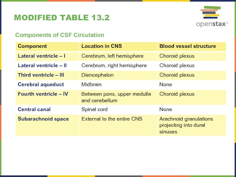
- 22. Modified table 13.2Components of CSF Circulation
- 23. 13.4 The Peripheral Nervous System Major section
- 24. Figure 13.19Dorsal Root GanglionThe cell bodies of
- 25. Figure 13.20Spinal Cord and Root GanglionThe slide
- 26. Figure 13.21Nerve StructureThe structure of a nerve
- 27. Figure 13.22Close-Up of Nerve TrunkZoom in on
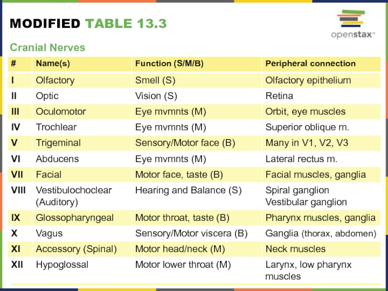
- 28. Figure 13.23The Cranial NervesThe anatomical arrangement of
- 29. Modified table 13.3Cranial Nerves
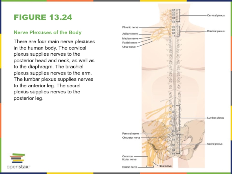
- 30. Figure 13.24Nerve Plexuses of the BodyThere are
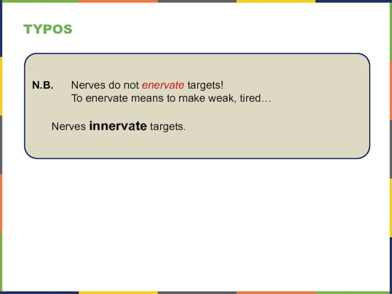
- 31. TyposN.B. Nerves do not enervate targets! To enervate means to make weak, tired… Nerves innervate targets.
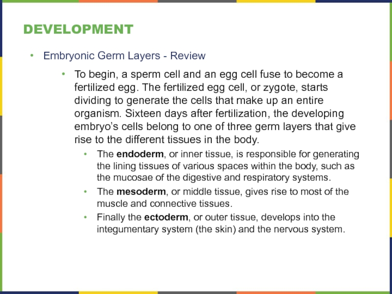
- 32. developmentEmbryonic Germ Layers - ReviewTo begin, a
- 33. developmentNeural Tube FormationEarly formation of the nervous
- 34. agingAnosmiaThe sensory neurons of the olfactory epithelium
- 35. everyday connectionsLeft Brain/Right BrainTo say that people
- 36. disorders & homeostatic imbalancesSpina BifidaResults from a
- 37. Figure 13.5Spinal BifidaSpina bifida is a birth
- 38. disorders & homeostatic imbalancesBasal Nuclei and Parkinson’s
- 39. disorders & homeostatic imbalancesMeningitisMeningitis is an inflammation
- 40. disorders & homeostatic imbalancesCNS Perfusion DisordersWithout a
- 41. interactive linksWatch this animation http://openstaxcollege.org/l/braindevel to examine
- 42. interactive linksWatch this animation http://openstaxcollege.org/l/bloodflow1 to see
- 43. interactive linksVisit this site http://openstaxcollege.org/l/NYTmeningitis to read
- 44. Errors in CHAPTER ReviewThis PowerPoint presentation is
- 45. Скачать презентанцию
Слайды и текст этой презентации
Слайд 2MAJOR CHAPTER Objectives
Relate the developmental processes of the embryonic nervous
system to the adult structures
Name the major regions of the
adult nervous systemLocate regions of the cerebral cortex on the basis of anatomical landmarks common to all human brains
Describe the regions of the spinal cord in cross-section
List the cranial nerves in order of anatomical location and provide the central and peripheral connections
List the spinal nerves by vertebral region and by which nerve plexus each supplies
Add:
Explain and illustrate the concept of somatotopy (associated to that of “localization of function”)
Be able to discuss normal development and selected aging issues
Be able to discuss selected, associated disorders
Слайд 313.1 The Embryologic Perspective
Major section Objectives
Describe the growth and differentiation
of the neural tube
Relate the different stages of development to
the adult structures of the central nervous systemExplain the expansion of the ventricular system of the adult brain from the central canal of the neural tube
Describe the connections of the diencephalon and cerebellum on the basis of patterns of embryonic development
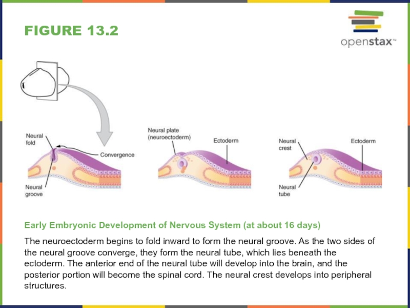
Слайд 4Figure 13.2
Early Embryonic Development of Nervous System (at about 16
days)
The neuroectoderm begins to fold inward to form the neural
groove. As the two sides of the neural groove converge, they form the neural tube, which lies beneath the ectoderm. The anterior end of the neural tube will develop into the brain, and the posterior portion will become the spinal cord. The neural crest develops into peripheral structures.Слайд 5Figure 13.3
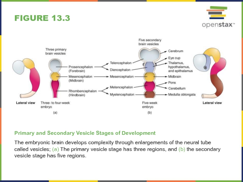
Primary and Secondary Vesicle Stages of Development
The embryonic brain
develops complexity through enlargements of the neural tube called vesicles;
(a) The primary vesicle stage has three regions, and (b) the secondary vesicle stage has five regions.Слайд 6Figure 13.4
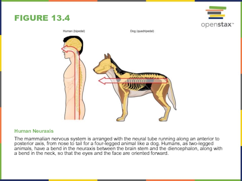
Human Neuraxis
The mammalian nervous system is arranged with the
neural tube running along an anterior to posterior axis, from
nose to tail for a four-legged animal like a dog. Humans, as two-legged animals, have a bend in the neuraxis between the brain stem and the diencephalon, along with a bend in the neck, so that the eyes and the face are oriented forward.Слайд 7table 13.1
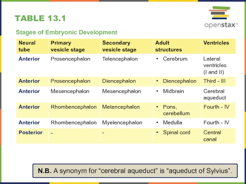
Stages of Embryonic Development
N.B. A synonym for “cerebral
aqueduct” is “aqueduct of Sylvius”.
Слайд 813.2 The Central Nervous System
Major section Objectives
Name the major regions
of the adult brain
Describe the connections between the cerebrum and
brain stem through the diencephalon, and from those regions into the spinal cordRecognize the complex connections within the subcortical structures of the basal nuclei
Explain the arrangement of gray and white matter in the spinal cord
Слайд 9Figure 13.6
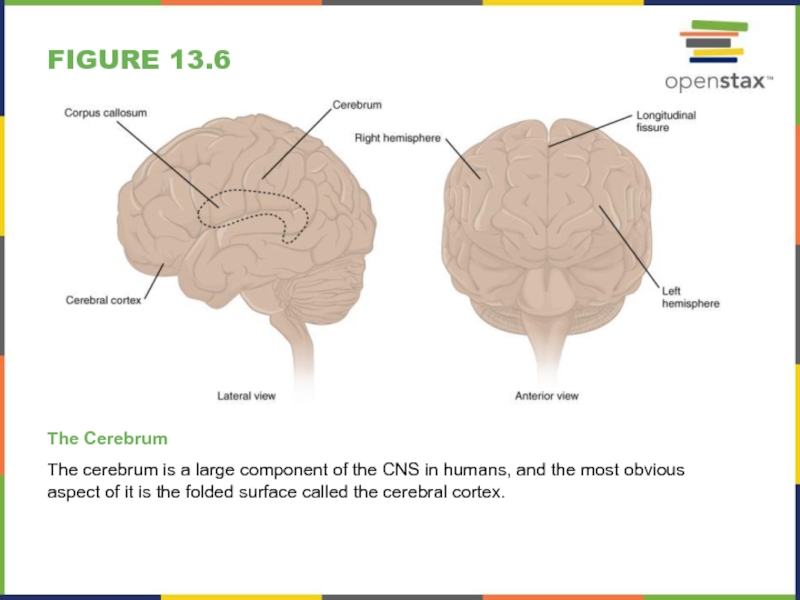
The Cerebrum
The cerebrum is a large component of
the CNS in humans, and the most obvious aspect of
it is the folded surface called the cerebral cortex.Слайд 10Figure 13.7
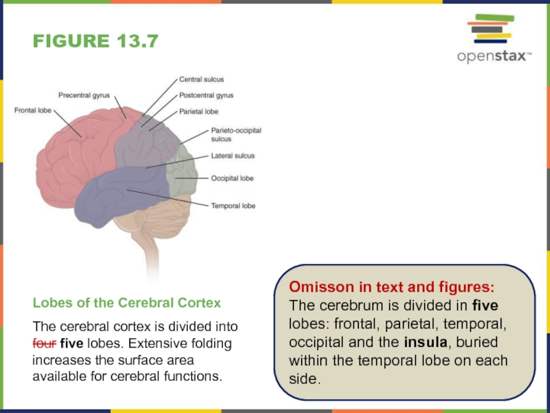
Lobes of the Cerebral Cortex
The cerebral cortex is divided
into four five lobes. Extensive folding increases the surface area
available for cerebral functions.Omisson in text and figures: The cerebrum is divided in five lobes: frontal, parietal, temporal, occipital and the insula, buried within the temporal lobe on each side.
Слайд 11Figure 13.8
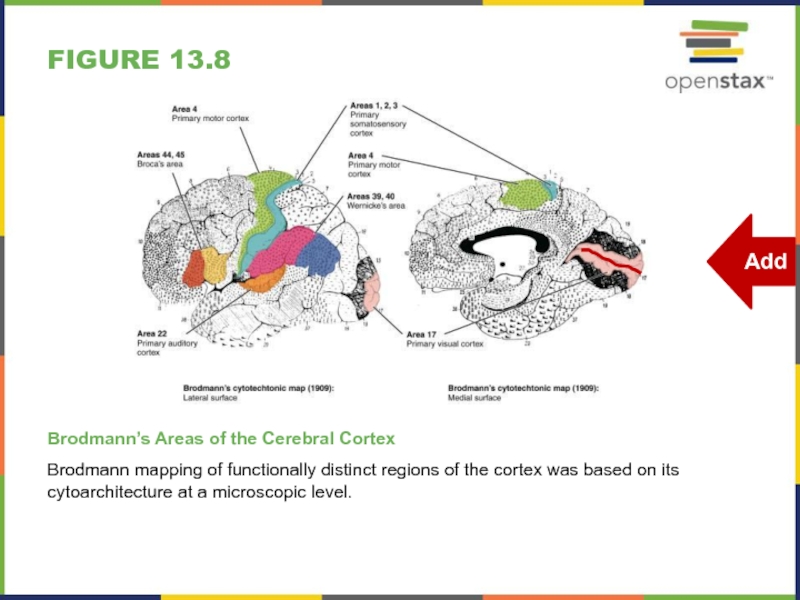
Brodmann’s Areas of the Cerebral Cortex
Brodmann mapping of functionally
distinct regions of the cortex was based on its cytoarchitecture
at a microscopic level.Слайд 12Figure 13.9
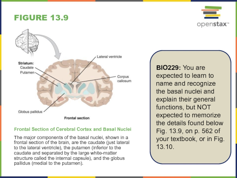
Frontal Section of Cerebral Cortex and Basal Nuclei
The major
components of the basal nuclei, shown in a frontal section
of the brain, are the caudate (just lateral to the lateral ventricle), the putamen (inferior to the caudate and separated by the large white-matter structure called the internal capsule), and the globus pallidus (medial to the putamen).BIO229: You are expected to learn to name and recognize the basal nuclei and explain their general functions, but NOT expected to memorize the details found below Fig. 13.9, on p. 562 of your textbook, or in Fig. 13.10.
Слайд 13Figure 13.11
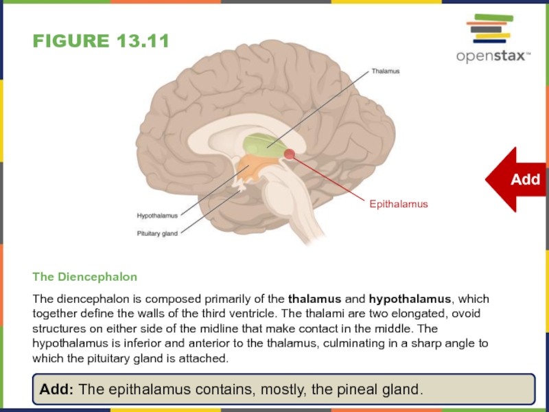
The Diencephalon
The diencephalon is composed primarily of the thalamus
and hypothalamus, which together define the walls of the third
ventricle. The thalami are two elongated, ovoid structures on either side of the midline that make contact in the middle. The hypothalamus is inferior and anterior to the thalamus, culminating in a sharp angle to which the pituitary gland is attached.Add: The epithalamus contains, mostly, the pineal gland.
Слайд 14Figure 13.12
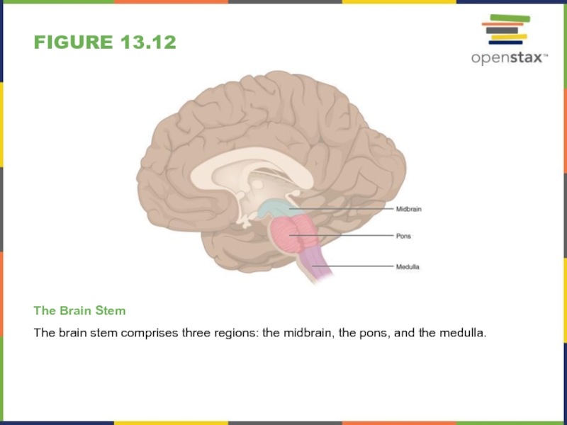
The Brain Stem
The brain stem comprises three regions: the
midbrain, the pons, and the medulla.
Слайд 15Figure 13.13
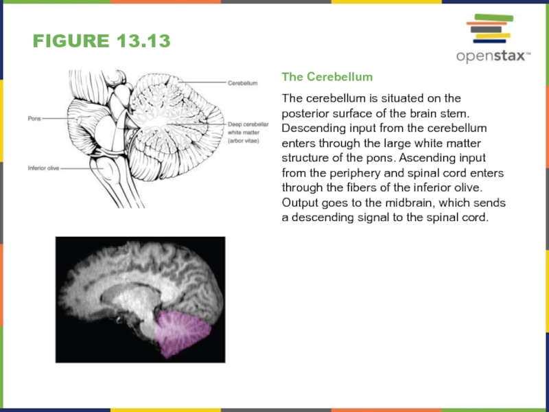
The Cerebellum
The cerebellum is situated on the posterior surface
of the brain stem. Descending input from the cerebellum enters
through the large white matter structure of the pons. Ascending input from the periphery and spinal cord enters through the fibers of the inferior olive. Output goes to the midbrain, which sends a descending signal to the spinal cord.Слайд 16Figure 13.14
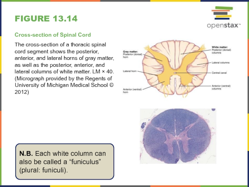
Cross-section of Spinal Cord
The cross-section of a thoracic spinal
cord segment shows the posterior, anterior, and lateral horns of
gray matter, as well as the posterior, anterior, and lateral columns of white matter. LM × 40. (Micrograph provided by the Regents of University of Michigan Medical School © 2012)N.B. Each white column can also be called a “funiculus” (plural: funiculi).
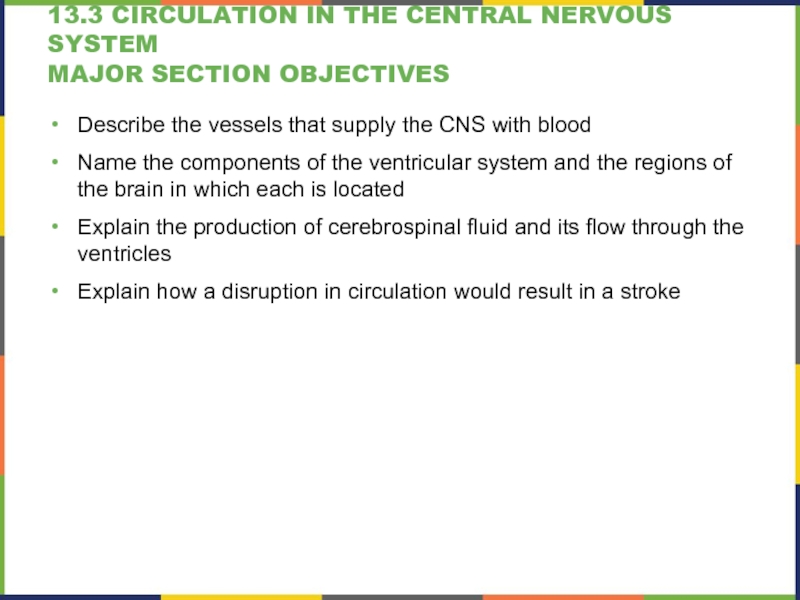
Слайд 1713.3 Circulation in the Central Nervous System
Major section Objectives
Describe the
vessels that supply the CNS with blood
Name the components of
the ventricular system and the regions of the brain in which each is locatedExplain the production of cerebrospinal fluid and its flow through the ventricles
Explain how a disruption in circulation would result in a stroke
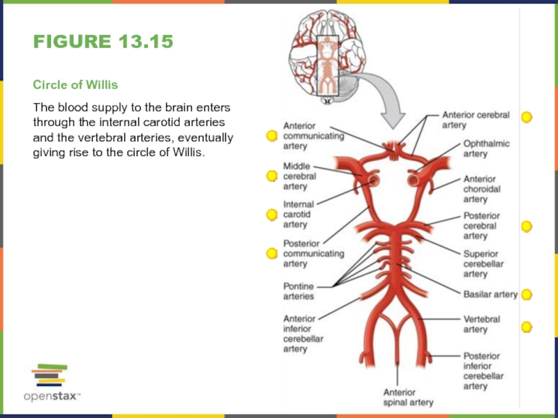
Слайд 18Figure 13.15
Circle of Willis
The blood supply to the brain enters
through the internal carotid arteries and the vertebral arteries, eventually
giving rise to the circle of Willis.Слайд 19Figure 13.16
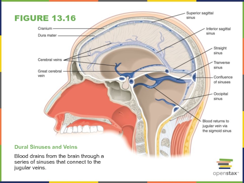
Dural Sinuses and Veins
Blood drains from the brain through
a series of sinuses that connect to the jugular veins.
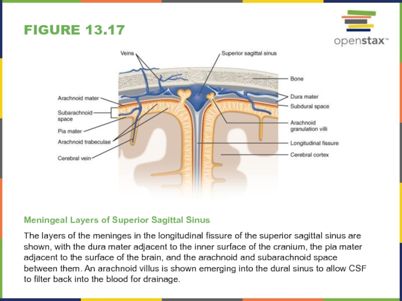
Слайд 20Figure 13.17
Meningeal Layers of Superior Sagittal Sinus
The layers of the
meninges in the longitudinal fissure of the superior sagittal sinus
are shown, with the dura mater adjacent to the inner surface of the cranium, the pia mater adjacent to the surface of the brain, and the arachnoid and subarachnoid space between them. An arachnoid villus is shown emerging into the dural sinus to allow CSF to filter back into the blood for drainage.Слайд 21Figure 13.18
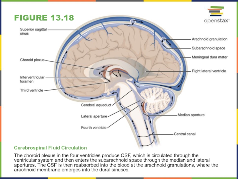
Cerebrospinal Fluid Circulation
The choroid plexus in the four ventricles
produce CSF, which is circulated through the ventricular system and
then enters the subarachnoid space through the median and lateral apertures. The CSF is then reabsorbed into the blood at the arachnoid granulations, where the arachnoid membrane emerges into the dural sinuses.Слайд 2313.4 The Peripheral Nervous System
Major section Objectives
Describe the structures found
in the PNS
Distinguish between somatic and autonomic structures, including the
special peripheral structures of the enteric nervous systemName the twelve cranial nerves and explain the functions associated with each
Describe the sensory and motor components of spinal nerves and the plexuses that they pass through
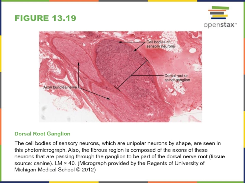
Слайд 24Figure 13.19
Dorsal Root Ganglion
The cell bodies of sensory neurons, which
are unipolar neurons by shape, are seen in this photomicrograph.
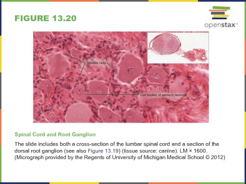
Also, the fibrous region is composed of the axons of these neurons that are passing through the ganglion to be part of the dorsal nerve root (tissue source: canine). LM × 40. (Micrograph provided by the Regents of University of Michigan Medical School © 2012)Слайд 25Figure 13.20
Spinal Cord and Root Ganglion
The slide includes both a
cross-section of the lumbar spinal cord and a section of
the dorsal root ganglion (see also Figure 13.19) (tissue source: canine). LM × 1600. (Micrograph provided by the Regents of University of Michigan Medical School © 2012)Слайд 26Figure 13.21
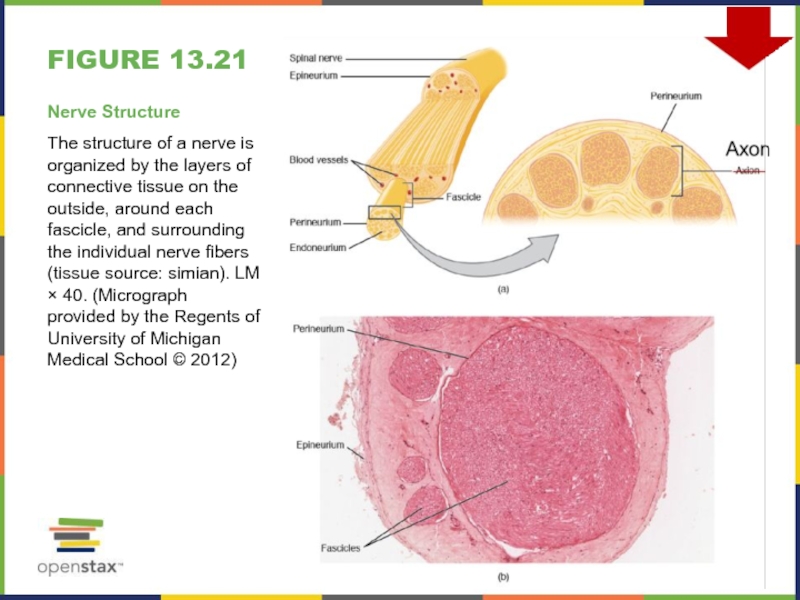
Nerve Structure
The structure of a nerve is organized by
the layers of connective tissue on the outside, around each
fascicle, and surrounding the individual nerve fibers (tissue source: simian). LM × 40. (Micrograph provided by the Regents of University of Michigan Medical School © 2012)Слайд 27Figure 13.22
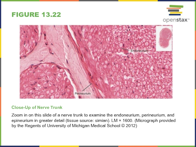
Close-Up of Nerve Trunk
Zoom in on this slide of
a nerve trunk to examine the endoneurium, perineurium, and epineurium
in greater detail (tissue source: simian). LM × 1600. (Micrograph provided by the Regents of University of Michigan Medical School © 2012)Слайд 28Figure 13.23
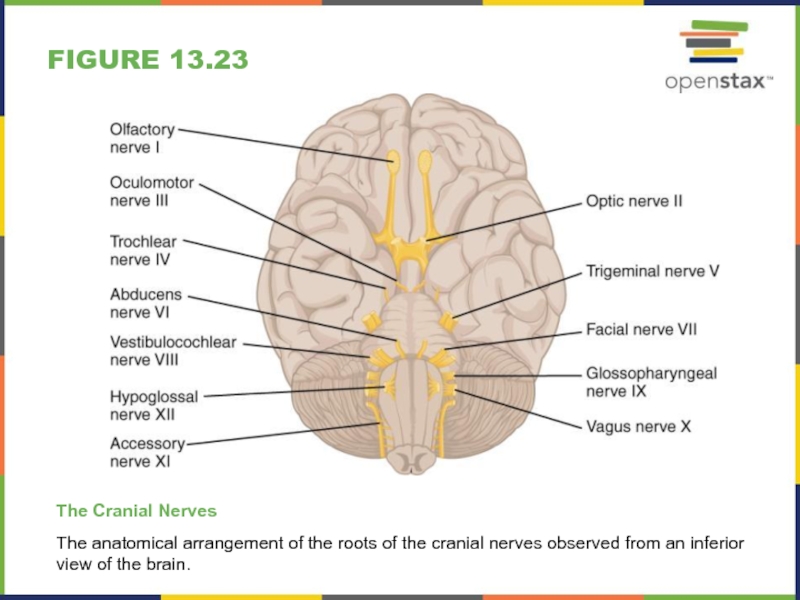
The Cranial Nerves
The anatomical arrangement of the roots of
the cranial nerves observed from an inferior view of the
brain.Слайд 30Figure 13.24
Nerve Plexuses of the Body
There are four main nerve
plexuses in the human body. The cervical plexus supplies nerves
to the posterior head and neck, as well as to the diaphragm. The brachial plexus supplies nerves to the arm. The lumbar plexus supplies nerves to the anterior leg. The sacral plexus supplies nerves to the posterior leg.Слайд 31Typos
N.B. Nerves do not enervate targets!
To enervate means to make
weak, tired…
Nerves innervate targets.
Слайд 32development
Embryonic Germ Layers - Review
To begin, a sperm cell and
an egg cell fuse to become a fertilized egg. The
fertilized egg cell, or zygote, starts dividing to generate the cells that make up an entire organism. Sixteen days after fertilization, the developing embryo’s cells belong to one of three germ layers that give rise to the different tissues in the body.The endoderm, or inner tissue, is responsible for generating the lining tissues of various spaces within the body, such as the mucosae of the digestive and respiratory systems.
The mesoderm, or middle tissue, gives rise to most of the muscle and connective tissues.
Finally the ectoderm, or outer tissue, develops into the integumentary system (the skin) and the nervous system.
Слайд 33development
Neural Tube Formation
Early formation of the nervous system depends on
the formation of the neural tube. A groove forms along
the dorsal surface of the embryo, which becomes deeper until its edges meet and close off to form the tube. If this fails to happen, especially in the posterior region where the spinal cord forms, a developmental defect called spina bifida occurs. The closing of the neural tube is important for more than just the proper formation of the nervous system. The surrounding tissues are dependent on the correct development of the tube. The connective tissues surrounding the CNS can be involved as well.Слайд 34aging
Anosmia
The sensory neurons of the olfactory epithelium have a limited
lifespan of approximately one to four months, and new ones
are made on a regular basis. The new neurons extend their axons into the CNS by growing along the existing fibers of the olfactory nerve.Anosmia is the loss of the sense of smell.
Anosmia is often the result of the olfactory nerve being severed, usually because of blunt force trauma to the head. Following the damage, new neurons cannot regrow without intact fibers to follow.
The ability of these neurons to be replaced is also lost with age. Age-related anosmia is hence a slow loss of the sensory neurons responsible for olfaction, with no new neurons born to replace them.
Слайд 35everyday connections
Left Brain/Right Brain
To say that people are “right-brained” or
“left-brained” is an oversimplification of an important concept about the
cerebral hemispheres.There is some lateralization of function, in which the left side of the brain is devoted to language function and the right side is devoted to spatial and nonverbal reasoning in most individuals.
But whereas these functions are predominantly associated with those sides of the brain, there is no monopoly by either side on these functions. Many pervasive functions, such as language, are distributed globally around the cerebrum.
The term “cerebral dominance” is nonetheless still used to describe the side of the cerebrum where the language of Broca and Wernicke are found (nothing to do with being right- or left-handed!).
Слайд 36disorders & homeostatic imbalances
Spina Bifida
Results from a failure of the
neural tube to close.Two forms present with an external cyst,
this mass forming a “second head” that gave this condition its name (bi-fida = two heads).There are three classes of this disorder: occulta, meningocele, and myelomeningocele.
Spina bifida occulta, is the mildest because the vertebral bones do not fully surround the spinal cord, but the spinal cord itself is not affected. No functional differences may be noticed, which is what the word occulta means; it is hidden spina bifida.
The other two types both involve the formation of a cyst—a fluid-filled sac of the connective tissues that cover the spinal cord called the meninges. Surgery to close the opening or to remove the cyst is necessary; the earlier that it can be performed, the better the chances of controlling or limiting further damage or infection at the opening.
“Meningocele” means that the meninges protrude through the spinal column but nerves may not be involved and few symptoms are present, though complications may arise later in life.
“Myelomeningocele” means that the meninges protrude and spinal nerves are involved, and therefore severe neurological symptoms can be present.
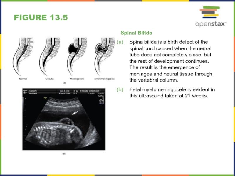
Слайд 37Figure 13.5
Spinal Bifida
Spina bifida is a birth defect of the
spinal cord caused when the neural tube does not completely
close, but the rest of development continues. The result is the emergence of meninges and neural tissue through the vertebral column.Fetal myelomeningocele is evident in this ultrasound taken at 21 weeks.
Слайд 38disorders & homeostatic imbalances
Basal Nuclei and Parkinson’s Disease
Parkinson’s disease is
a disorder of the basal nuclei, specifically of the substantia
nigra. As the dopamine-producing neurons in the substantia nigra pars compacta die, they fail to modulate the activity of the basal nuclei properly – causing too much inhibition of cortical activity which results in a hypokinetic disorder.Parkinson’s disease is neurodegenerative, meaning that neurons die that cannot be replaced, so there is no cure for the disorder.
Treatments for Parkinson’s disease are aimed at increasing dopamine levels among basal nuclei by providing the amino acid L-DOPA, which is a precursor to the neurotransmitter dopamine that can cross the blood-brain barrier (dopamine cannot).
Unfortunately, patients become less responsive to L-DOPA treatment as time progresses - and the increased dopamine levels created elsewhere in the brain can be associated with psychosis or schizophrenia.
Слайд 39disorders & homeostatic imbalances
Meningitis
Meningitis is an inflammation of the meninges,
the three layers of fibrous membrane that surround the CNS,
and can be caused by infection by bacteria or viruses. The particular pathogens are not special to meningitis.Symptoms of meningitis include: fever, chills, nausea, vomiting, light sensitivity, soreness of the neck, or severe headache, but also changes in mental state (confusion, memory deficits, etc.).
The primary test for meningitis is a lumbar puncture.
Viral meningitis is usually the result of common enteroviruses (such as those that cause intestinal disorders), but may be the result of the herpes virus or West Nile virus. Viral meningitis cannot be treated with antibiotics but fortunately, viral forms tend to be milder.
Bacterial meningitis can be caused by Streptococcus or Staphylococcus, among many others. It is treated by antibiotics. Bacterial meningitis tends to be more severe, with fatality in 5 to 40 percent of children and 20 to 50 percent of adults.
Слайд 40disorders & homeostatic imbalances
CNS Perfusion Disorders
Without a steady supply of
oxygen, and to a lesser extent glucose, the nervous tissue
in the brain cannot keep up its extensive electrical activity. These nutrients get into the brain through the blood, and if blood flow is interrupted, neurological function is compromised.A stroke (disruption of blood supply to the brain) is caused by a blockage to an artery from some type of embolus: a blood clot, a fat embolus, or an air bubble.
When the blood cannot travel through the artery, the surrounding tissue that is deprived starves and dies. Strokes will often result in the loss of very specific functions.
Related to strokes are transient ischemic attacks (TIAs) - “mini-strokes” - in which a physical blockage may temporarily affect a region. Lost neurological function may return.
Sometimes, treatment with blood-thinning drugs can alleviate the problem, and recovery is possible.
Because the nervous system is adaptable, even with neuron death, victims of strokes can recover, or more accurately relearn, some functions with physical, occupational, and speech therapy.
Слайд 41interactive links
Watch this animation http://openstaxcollege.org/l/braindevel to examine the development of
the brain, starting with the neural tube.
Watch this video http://openstaxcollege.org/l/whitematter
to learn about the white matter in the cerebrum that develops during childhood and adolescence.FYI only BIO229: The videos http://openstaxcollege.org/l/basalnuclei1 and http://openstaxcollege.org/l/basalnuclei2 describe two basal nuclei-related pathways that process information within the cerebrum.
Watch this video http://openstaxcollege.org/l/graymatter to learn about the gray matter of the spinal cord that receives input from fibers of the dorsal (posterior) root and sends information out through the fibers of the ventral (anterior) root.
Visit this site http://openstaxcollege.org/l/parkinsons for a thorough explanation of Parkinson’s disease.
Read this article http://openstaxcollege.org/l/hugebrain in which the author explores the current understanding of what might have happened to increase the size of the human brain relative to that of the chimpanzee.
Слайд 42interactive links
Watch this animation http://openstaxcollege.org/l/bloodflow1 to see how blood flows
to the brain and passes through the circle of Willis
before being distributed through the cerebrum.Watch this video http://openstaxcollege.org/l/lumbarpuncture that describes the procedure known as the lumbar puncture, a medical procedure used to sample the CSF.
Watch this animation http://openstaxcollege.org/l/CSFflow that shows the flow of CSF through the brain and spinal cord, and how it originates from the ventricles and then spreads into the space within the meninges, where the fluids then move into the venous sinuses to return to the cardiovascular circulation.
Explore a section of spinal cord and a dorsal root ganglion at http://openstaxcollege.org/l/spinalroot.
Explore a section of nerve trunk at http://openstaxcollege.org/l/nervetrunk.
