Разделы презентаций
- Разное
- Английский язык
- Астрономия
- Алгебра
- Биология
- География
- Геометрия
- Детские презентации
- Информатика
- История
- Литература
- Математика
- Медицина
- Менеджмент
- Музыка
- МХК
- Немецкий язык
- ОБЖ
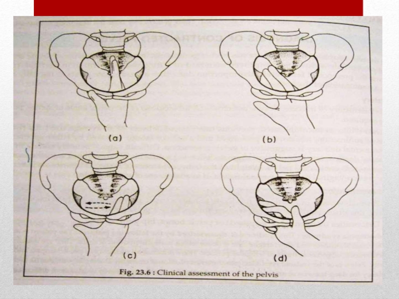
- Обществознание
- Окружающий мир
- Педагогика
- Русский язык
- Технология
- Физика
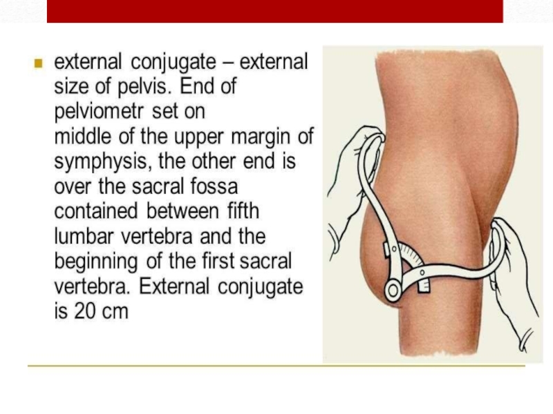
- Философия
- Химия
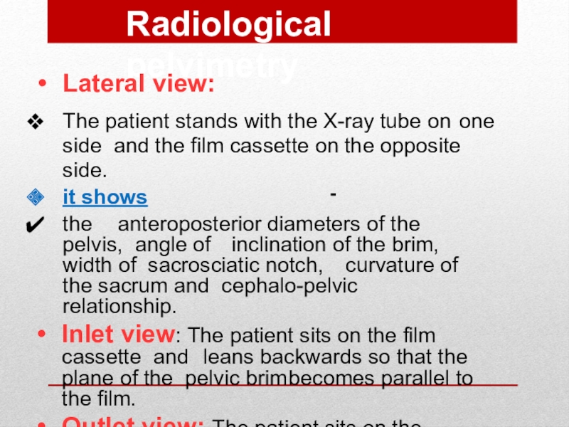
- Шаблоны, картинки для презентаций
- Экология
- Экономика
- Юриспруденция
CEPHALO PELVIC DISPROPORTION (CPD)
Содержание
- 1. CEPHALO PELVIC DISPROPORTION (CPD)
- 2. Слайд 2
- 3. CPD either due to :-The baby’s head
- 4. Causes :-Large baby due to:Hereditary factorsDiabetesPostmaturity (still
- 5. Contracted Pelvis
- 6. Contracted PelvisDefinition:Anatomical definition: It is a pelvis
- 7. Factors influencing the size and shapeof the pelvis:Developmental
- 8. Etiology of Contracted PelvisCauses in the pelvisDevelopmental
- 9. Naegele’s pelvis: absence of one sacral alaRobert’s
- 10. Causes in the pelvisMetabolic:Rickets.Osteomalacia (triradiate pelvic brim).Traumatic: as fractures.Neoplastic: as osteoma.Infection : TBEtiology of ContractedPelvis
- 11. Causes in the spineLumbarkyphosisLumbar scoliosisSpondylolisthesis:The 5th lumbar
- 12. Causes in the lower limbsDislocation of one
- 13. PelvisHistoryRickets: is expected if there is a
- 14. ExaminationGeneral examination:Gait: abnormal gait suggesting abnormalities in
- 15. Examinat ionGeneral examination:Manifestations of rickets as:square headrosary
- 16. Abdominal examination:Nonengagement of the head:in the last 3-4 weeks in primigravida.Pendulous abdomen:in a primigravida.Malpresentations:are more common.Pelvis
- 17. Pelvimetry :It is assessment of the pelvic diameters
- 18. Pelvimetry:2.Imaging pelvimetry:X-ray.Computed tomography (CT).Magnetic resonance imaging (MRI)
- 19. Internal pelvimetryis done through vaginalexamination1. The inlet:Palpation of
- 20. Internal pelvimetry2.The cavity:Height, thickness and inclination of
- 21. 2.The cavity:d.Ischial spines:Whether it is blunt (difficult
- 22. 2.The cavity:e.Interspinous diameter: By using the 2
- 23. 3- The outlet:Subpubic angle: Normally, it admits
- 24. Слайд 24
- 25. External pelvimetryThom’s, Jarcho’s or crossing pelvimeter can
- 26. Слайд 26
- 27. Radiological pelvimetryLateral view:The patient stands with the X-ray
- 28. CephalometryUltrasonography: is the safe accurate and easy
- 29. Cephalopelvic disproportion testsThese are done to detect
- 30. (2) Muller - Kerr’s method:It is more
- 31. Degrees of DisproportionMinor disproportion:The anterior surface of
- 32. Degrees of Contracted PelvisMinor degree: The true
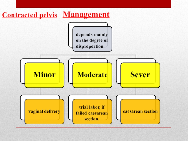
- 33. Managementdepends mainly on the degree of disproportionMinorvaginal deliveryModeratetrial labor, if failed caesarean section.Severcaesarean sectionContracted pelvis
- 34. Trial of LabourIt is a clinical test
- 35. Procedure :Trial is carried out in a
- 36. Indications of trial of labour:Young primigravida of good health.Moderate disproportion.Vertex presentation.No contracted outletAverage sized baby.Vertex presentation
- 37. Termination of trial of labour:Vaginal delivery: either
- 38. Indications of caesarean section in contracted pelvisModerate disproportion if
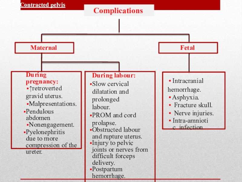
- 39. ComplicationsMaternalFetalContracted pelvis
- 40. Maternal:During pregnancy:Incarcerated retroverted gravid uterus.Malpresentations.Pendulous abdomen.Nonengagement.Pyelonephritis especial
- 41. Complications of Contracted PelvisDuring labour:Inertia, slow cervical
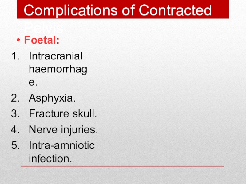
- 42. Foetal:Intracranial haemorrhage.Asphyxia.Fracture skull.Nerve injuries.Intra-amniotic infection.Complications of Contracted Pelvis
- 43. Слайд 43
- 44. Скачать презентанцию
Слайды и текст этой презентации
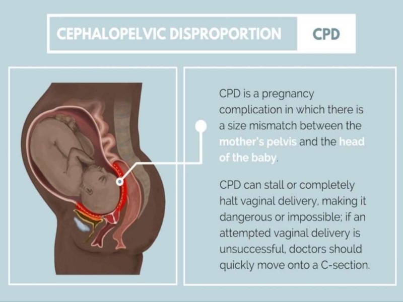
Слайд 1CEPHALO PELVIC DISPROPORTION
(CPD)
Teacher : Kamilova Irina Kaharovna
By: Sulur PerumalSwamy Venkatesh
Prabhu
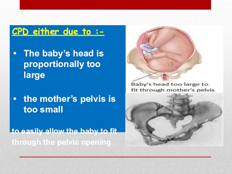
Слайд 3CPD either due to :-
The baby’s head is proportionally too
large
the mother’s pelvis is too small
to easily allow the baby
to fitthrough the pelvic opening.
Слайд 4Causes :-
Large baby due to:
Hereditary factors
Diabetes
Postmaturity (still pregnant after due
date has passed)
Multiparity (not the first pregnancy)
Abnormal fetal positions
contracted pelvis
Abnormally
shaped pelvisСлайд 6Contracted Pelvis
Definition:
Anatomical definition: It is a pelvis in which one
or more of its diameters is reduced below the normal
by one or more centimeters.Obstetric definition: It is a pelvis in which its size & shape is sufficiently abnormal that interfere with vaginal delivery of normal size fetus
Слайд 7Factors influencing the size and shape
of the pelvis:
Developmental factor: hereditary or
congenital.
Racial factor.
Nutritional factor: malnutrition results in small pelvis.
Sexual factor: as excessive
androgen may produce android pelvis.Metabolic factor: as rickets and osteomalacia.
Trauma, diseases or tumours of the bony pelvis, legs or spines.
Слайд 8Etiology of Contracted Pelvis
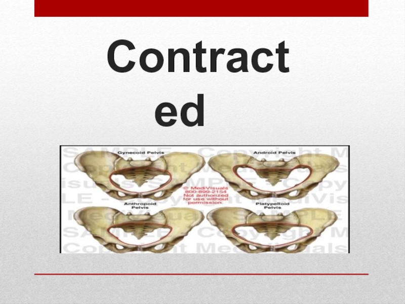
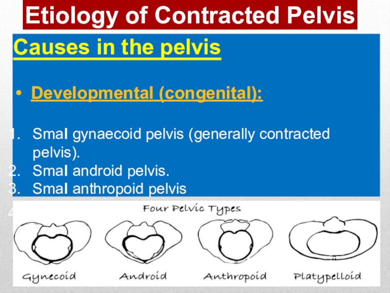
Causes in the pelvis
Developmental (congenital):
Smal gynaecoid pelvis
(generally contracted pelvis).
Smal android pelvis.
Smal anthropoid pelvis
Smal platypelloid pelvis (simple
flat pelvis)Слайд 9Naegele’s pelvis: absence of one sacral ala
Robert’s pelvis: absence of
both sacral alae.
High assimilation pelvis: The sacrum is composed of
6 vertebrae.Low assimilation pelvis: The sacrum is composed of 4 vertebrae.
Split pelvis: splitted symphysis pubis
Слайд 10Causes in the pelvis
Metabolic:
Rickets.
Osteomalacia (triradiate pelvic brim).
Traumatic: as fractures.
Neoplastic: as
osteoma.
Infection : TB
Etiology of ContractedPelvis
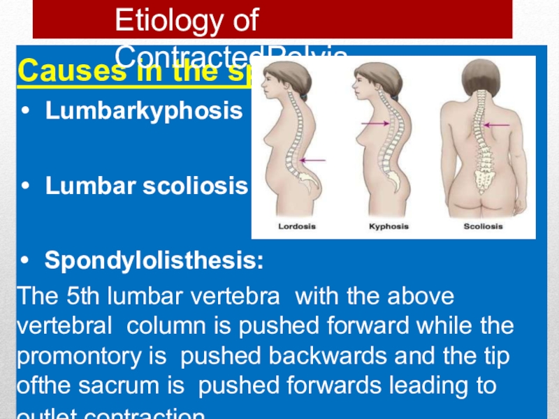
Слайд 11Causes in the spine
Lumbarkyphosis
Lumbar scoliosis
Spondylolisthesis:
The 5th lumbar vertebra with the above
vertebral column is pushed forward while the promontory is pushed
backwards and the tip ofthe sacrum is pushed forwards leading to outlet contraction.Etiology of ContractedPelvis
Слайд 12Causes in the lower limbs
Dislocation of one or bothfemurs.
Atrophy ofone
or both lower limbs.
N.B. oblique or asymmetric pelvis: one oblique
diameter is obviously shorter than theother. This can be found in:Diseases, fracture or tumours affecting one side.
Etiology of Contracted Pelvis
Слайд 13Pelvis
History
Rickets: is expected if there is a history of delayed
walking and dentition.
Trauma or diseases: of the pelvis, spines or
lower limbs.Bad obstetric history: e.g. prolonged labour ended by:
difficult forceps
caesarean section or
still birth.
Слайд 14Examination
General examination:
Gait: abnormal gait suggesting abnormalities in the pelvis, spines
or lower limbs.
Height: women with less than 150 cmheight usual
y have contracted pelvis.Spines and lower limbs: may have a disease or lesion.( kyphosis,…)
Pelvis
Слайд 15Examinat ion
General examination:
Manifestations of rickets as:
square head
rosary beads in the
costalridges.
pigeon chest
Harrison’s sulcus and bow legs.
Dystocia dystrophia syndrome: the woman
is*short,obese stocky, subfertile, has android pelvis and
Pelvis
Слайд 16Abdominal examination:
Nonengagement of the head:
in the last 3-4 weeks in primigravida.
Pendulous
abdomen:
in a primigravida.
Malpresentations:
are more common.
Pelvis
Слайд 17Pelvimetry :
It is assessment of the pelvic diameters and capacity done
at 38-39 weeks.It includes:
1. Clinical pelvimetry:
Internal pelvimetry for:
inlet
cavity, and
outlet.
External pelvimetry for:
inlet and
outlet.
Pelvis
Слайд 18Pelvimetry:
2.Imaging pelvimetry:
X-ray.
Computed tomography (CT).
Magnetic resonance imaging (MRI) .
N.B. CTand MRI
are recent and accurate but expensive and not always available
so they are not in common use.Diagnosis of Contracted Pelvis
Слайд 19Internal pelvimetry
is done through vaginalexamination
1. The inlet:
Palpation of the forepelvis (pelvicbrim):
The
index and middle fingers are moved along the pelvic brim.
Note whether it is round or angulated, causing the fingers to dip into a V- shaped depression behind the symphysis.Diagonal conjugate:
Try to palpate the sacral promontory to measure the diagonal conjugate. Normally, it is
12.5 cm and cannot be reached. If it is felt the pelvis is considered contracted and the true conjugate can be calculated by subtracting 1.5 cm from the diagonal conjugate .This assessment is not done if the head isengaged.
Слайд 20Internal pelvimetry
2.The cavity:
Height, thickness and inclination of the symphysis.
Shape and
inclination of the sacrum.
Side walls: Todetermine whether it is straight,
convergent or divergent starting from the pelvic brim down to the base of ischial spines in the direction of the base of the ischial tuberosity. Then relation between the index and middle finger of the base of ischial spines and the thumb of the other hand on the ischial tuberosity is detected. If the thumb is medial the side wall is convergent and if lateral it is divergent.Слайд 212.The cavity:
d.Ischial spines:
Whether it is blunt (difficult to identify at
all), prominent (easily felt but not large) or very prominent (large and
encroaching on the mid- plane).The ischial spines can be located by following the sacrospinous ligament to its
lateral end.
Internal pelvimetry
Слайд 222.The cavity:
e.Interspinous diameter: By using the 2 examining fingers, if
both spines can be touched simultaneously, the interspinous diameter is
£ 9.5 cm i.e. inadequate for an average-sized baby.f. Sacrosciatic notch: If the sacrospinous ligament is two and half fingers, the sacrosciatic notch is considered adequate.
Internal pelvimetry
Слайд 233- The outlet:
Subpubic angle: Normally, it admits 2fingers.
Mobility of the
coccyx: by pressing firmly on
it while an external hand on it
can determineits mobility.c.Anteroposterior diameter of the outlet: from the tip of the sacrum to the inferior edge of the symphysis.
Internal pelvimetry
Слайд 25External pelvimetry
Thom’s, Jarcho’s or crossing pelvimeter can be used for
external pelvimetry.
Interspinous diameter (25cm): between the anterior superior iliac spines.
Intercrestal
diameter (28 cm): between the most far points on the outer borders of the iliac crests.External conjugate (20 cm(.
Bituberous diameter (11cm)
Слайд 27Radiological pelvimetry
Lateral view:
The patient stands with the X-ray tube on one side
and the film cassette on the opposite side.
it shows
the anteroposterior diameters
of the pelvis, angle of inclination of the brim, width of sacrosciatic notch, curvature of the sacrum and cephalo-pelvic relationship.Inlet view: The patient sits on the film cassette and leans backwards so that the plane of the pelvic brim becomes parallel to the film.
Outlet view: The patient sits on the film cassette and leans forwards.
Слайд 28Cephalometry
Ultrasonography: is the safe accurate and easy method and can
detect:
The biparietal diameter (BPD)
The occipito-frontal diameter.
The circumference of the head.
Radiology
(X-ray: is difficult to interpret.Слайд 29Cephalopelvic disproportion tests
These are done to detect contracted inlet if
the head is not engaged in the last 3-4 weeks in
a primigravida.(1) Pinard’s method:
The patient evacuates her bladder and rectum.
The patient is placed in semi-sitting position to bring the foetal axis perpendicular tothe brim.
The left hand pushes the head downwards and backwards into the pelvis while the fingers of the right hand are put on the symphysis to detect disproportion.
Слайд 30(2) Muller - Kerr’s method:
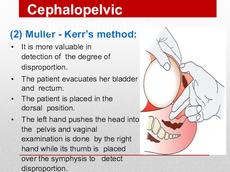
It is more valuable in detection
of the degree of disproportion.
The patient evacuates her bladder and rectum.
The
patient is placed in the dorsal position.The left hand pushes the head into the pelvis and vaginal examination is done by the right hand while its thumb is placed over the symphysis to detect disproportion.
Cephalopelvic disproportion tests
Слайд 31Degrees of Disproportion
Minor disproportion:
The anterior surface of the head is
in line with the posterior surface of the symphysis. During
labour the head is engaged due to moulding and vaginaldelivery can be achieved.Moderate disproportion 1st degree disproportion):The anterior surface of the head is in line with the anterior surface of the symphysis. Vaginal delivery may or may not occur.
Marked disproportion 2nd degreedisproportion):
The head overrides the anterior surface of the symphysis. Vaginal delivery cannot occur.
Слайд 32Degrees of Contracted Pelvis
Minor degree: The true conjugate is 9-10
cm. It corresponds to minor disproportion.
Moderate degree: The true conjugate is
8-9 cm.It corresponds to moderate disproportion.
Severe degree: The true conjugate is 6-8 cm. It corresponds to marked disproportion.
Extreme degree: The true conjugate is less than 6 cm. Vaginal delivery is impossible even after craniotomy asthe bimastoid diameter (7.5 cm) is not crushed.
Слайд 33Management
depends mainly on the degree of disproportion
Minor
vaginal delivery
Moderate
trial labor, if
failed caesarean section.
Sever
caesarean section
Contracted pelvis
Слайд 34Trial of Labour
It is a clinical test for the factors
that cannot be determined before start of labouras:
Efficiency of uterine
contractions.Moulding of the head.
Yielding of the pelvis and soft tissues.
Слайд 35Procedure :
Trial is carried out in a hospital where facilities
for C.S is available.
Adequate analgesia.
Nothing by mouth.
Avoid premature rupture of
membranes by:rest in bed,
avoid high enema,
minimise vaginal examinations.
The patient is left for 2 hours in the 2nd stage with good uterine contractions under close supervision to the mother and foetus
Слайд 36Indications of trial of labour:
Young primigravida of good health.
Moderate disproportion.
Vertex
presentation.
No contracted outlet
Average sized baby.
Vertex presentation
Слайд 37Termination of trial of labour:
Vaginal delivery: either spontaneously or by forceps
if the head is engaged.
Caesarean section if: failed trial of
labouri.e. the head did not engageor
complications occur during trial as
foetal distress or prolapsed pulsating cord before full cervical dilatation.
Слайд 38Indications of caesarean section in contracted pelvis
Moderate disproportion if trial of labour
is contraindicated or failed.
Marked disproportion.
Extreme disproportion whether the foetus is
living or dead.Contracted outlet.
Contracted pelvis with other indicationsas;
elderly primigravida,
malpresentations, or
placenta praevia.
Слайд 40Maternal:
During pregnancy:
Incarcerated retroverted gravid uterus.
Malpresentations.
Pendulous abdomen.
Nonengagement.
Pyelonephritis especial y in high
assimilation pelvis due to more compression of the ureter.
Complications of
Contracted PelvisСлайд 41Complications of Contracted Pelvis
During labour:
Inertia, slow cervical dilatation and prolonged labour.
Premature
rupture of membranes and cord prolapse.
Obstructed labour and rupture uterus.
Necrotic genito-urinary
fistula.Injury to pelvic joints or nerves from difficult forceps delivery.
Postpartum haemorrhage.

