Разделы презентаций
- Разное
- Английский язык
- Астрономия
- Алгебра
- Биология
- География
- Геометрия
- Детские презентации
- Информатика
- История
- Литература
- Математика
- Медицина
- Менеджмент
- Музыка
- МХК
- Немецкий язык
- ОБЖ
- Обществознание
- Окружающий мир
- Педагогика
- Русский язык
- Технология
- Физика
- Философия
- Химия
- Шаблоны, картинки для презентаций
- Экология
- Экономика
- Юриспруденция
The main methods of patient work-up
Содержание
- 1. The main methods of patient work-up
- 2. X-radiationX-rays radiation – is X-rays, which were
- 3. X-rays may be used for: Detecting pathology;Diagnosing
- 4. Radiation Oncology Radiation Therapy is the use
- 5. Computed tomographyComputed tomography (x-ray CT) is a technology
- 6. As x-ray CT is the most common
- 7. Слайд 7
- 8. Magnetic resonance imagingIs a medical technique used
- 9. Слайд 9
- 10. MRI has a wide range of applications
- 11. How MRI works? To perform a
- 12. EndoscopyAn endoscopy is the visual examination of
- 13. Types of Endoscopy arthoscopy—used for joints, with
- 14. Скачать презентанцию
X-radiationX-rays radiation – is X-rays, which were discovered by German physics Wilhelm Conrad Roentgen on November 8, 1895. These are rays, which differ from the rays of visible light by wavelength
Слайды и текст этой презентации
Слайд 3X-rays may be used for:
Detecting pathology;
Diagnosing of oral cavities;
As a
radiation therapy of cancer including palliation.
Слайд 4Radiation Oncology
Radiation Therapy is the use of high energy x-rays,
usually electronically produced, to treat cancer. The machine most commonly
used for such treatment is a Linear Accelerator.The aim of Radiation Therapy is to treat the area of the tumor, whilst preserving the surrounding normal tissues and important structures (e.g. spinal cord).
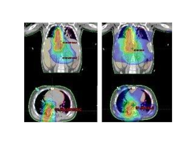
A course of Radiation Therapy consists of two parts. The first part is planning, where the doctor will decide the area to be treated. This is done in one appointment. The second part is treatment. A course of treatment is given on a daily basis from Monday to Friday and can consist of 1 single treatment or up to as many as 40 treatments. The number of treatments is determined by many factors. The type, size, and stage of the tumor are important as is the age and health of the patient. In between each treatment, the normal tissues have the ability to heal themselves, whilst the tumour cells do not.
Слайд 5Computed tomography
Computed tomography (x-ray CT) is a technology that uses computer-processed x-rays to
produce tomographic images of specific areas of the scanned object, allowing
the user to see what is inside it without cutting it open. Digital geometry processing is used to generate a three-dimensional image of the inside of an object from a large series of two-dimension alradiographic images taken around a single axis of rotation. Medical imaging is the most common application of x-ray CT. Its cross-sectional images are used for diagnostic and therapeutic purposes in various medical disciplines.The rest of this article discusses medical-imaging x-ray CT; industrial applications of x-ray CT are discussed at industrial computed tomography scanning.Слайд 6As x-ray CT is the most common form of CT
in medicine and various other contexts, the term computed tomography alone (or CT)
is often used to refer to x-ray CT, although other types exist (such as positron emission tomography[PET] and single-photon emission computed tomography 8Older and less preferred terms that also refer to x-ray CT are computed axial tomography (CAT scan) and computer-assisted tomography. X-ray CT is a form of radiography, although the word "radiography" used alone usually refers, by wide convention, to non-tomographic radiography.Слайд 8Magnetic resonance imaging
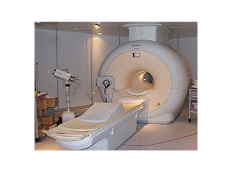
Is a medical technique used in radiology to
investigate the anatomy and function of the body in both
health and disease. MRI scanners use strong magnetic fields and radiowaves to form images of the body. The technique is widely used in hospitals for medical diagnosis, staging of disease and for follow-up without exposure to ionizing radiation.Слайд 10MRI has a wide range of applications in medical diagnosis
and there are estimated to be over 25,000 scanners in
use worldwide. MRI has an impact on diagnosis and treatment in many specialties although the effect on improved health outcomes is uncertain. Since MRI does not use any ionizing radiation its use is recommended in preference to CT when either modality could yield the same information. MRI is in general a safe technique but the number of incidents causing patient harm have risen.Contraindications to MRI include most cochlear implants and cardiac pacemakers, shrapnel and metallic foreign bodies in the orbits, and some ferromagnetic surgical implants. The safety of MRI during the first trimester of pregnancy is uncertain, but it may be preferable to alternative options. The sustained increase in demand for MRI within the healthcare industry has led to concerns about cost effectiveness and over diagnosis.Слайд 11How MRI works?
To perform a study the patient is positioned
within an MRI scanner which forms a strong magnetic field
around the area to be imaged. Most medical applications rely on detecting a radio frequency signal emitted by excited hydrogen atoms in the body (present in any tissue containing water molecules) using energy from an oscillating magnetic field applied at the appropriate resonant frequency. The orientation of the image is controlled by varying the main magnetic field using gradient coils. As these coils are rapidly switched on and off they create the characteristic repetitive noises of an MRI scan. The contrast between different tissues is determined by the rate at which excited atoms return to the equilibrium state. Exogenous contrast agents may be given intravenously, orally or intra-articularly.MRI requires a magnetic field that is both strong and uniform. The field strength of the magnet is measured in tesla – and while the majority of systems operate at 1.5T commercial systems are available between 0.2T–7T. Most clinical magnets are superconducting which requires liquid helium. The lower field strengths can be achieved with permanent magnets, which are often used in "open" MRI scanners for claustrophobic patients.
Слайд 12Endoscopy
An endoscopy is the visual examination of the interior of
a hollow body organ by use of an endoscope. An
endoscope is an instrument used to examine the interior of a hollow organ or cavity of the body done by inserting it directly into the organ. Endoscopy can also refer to using a borescope in technical situations where direct line of-sight observationСлайд 13Types of Endoscopy
arthoscopy—used for joints, with the scope inserted through
a small incision near the joint being examined
bronchoscopy—used for the
lungs, with the scope inserted into your nose or your mouthcolonoscopy—used for the colon, with the scope inserted through the anus
cystoscopy—used for the bladder, with the scope going through the urethra (the hole where urine is expelled)
enteroscopy—used for the small intestine, with the scope going through the mouth or anus
hysteroscopy—used for the inside of the uterus, with the scope inserted through the vagina
laparoscopy—used for the abdominal or pelvic area, with the scope inserted through a small incision near the area that is being examined
laryngoscopy—used for the voice box (larynx), with the scope inserted through the mouth or nostril
mediastinoscopy—used for the area between the lungs, with the scope inserted through an incision above the breastbone
upper gastrointestinal endoscopy—used for the esophagus and upper intestinal tract, with the scope inserted through the mouth
ureteroscopy—used to examine the ureter (the tube that carries urine from the kidneys to the bladder), with the scope inserted through the urethra (the tube that carries urine from the bladder out of the body).
