Разделы презентаций
- Разное
- Английский язык
- Астрономия
- Алгебра
- Биология
- География
- Геометрия
- Детские презентации
- Информатика
- История
- Литература
- Математика
- Медицина
- Менеджмент
- Музыка
- МХК
- Немецкий язык
- ОБЖ
- Обществознание
- Окружающий мир
- Педагогика
- Русский язык
- Технология
- Физика
- Философия
- Химия
- Шаблоны, картинки для презентаций
- Экология
- Экономика
- Юриспруденция
VOGT-KOYANAGI-HARADA DISEASE
Содержание
- 1. VOGT-KOYANAGI-HARADA DISEASE
- 2. Multisystem diseaseChronic, bilateral, granulomatous panuveitis associated with
- 3. VKH DiseaseIndividuals with a predisposing genetic background.Ethnic
- 4. VKH DiseaseGenetic background rather than degree of
- 5. VKH DiseaseRemains unknown.T-lymphocyte mediated autoimmunity directed against
- 6. VKH DiseaseTyrosinase family proteins are enzymes for
- 7. VKH DiseaseTh cells from peripheral blood of
- 8. VKH DiseaseIL-23 stimulated production of IL-17 by
- 9. VKH DiseaseVKH-like disease in patients treated with
- 10. VKH DiseaseGranulomatous panuveitis.Lymphocytes, epitheloid cells, few plasma
- 11. VKH DiseaseDalen-Fuchs’ nodules: Lymphocytes, epitheloid cells, pigment-laden
- 12. VKH DiseaseCertain racial groups.Immunogenetic predisposition.Strong association with
- 13. VKH DiseaseVKH disease in monozygotic twinsFamilial VKH
- 14. VKH DiseaseIntegumentary ManifestationsSensitivity of hair and skin
- 15. Clinical Features Extraocular ManifestationsVKH DiseaseNeurologic ManifestationsMost common
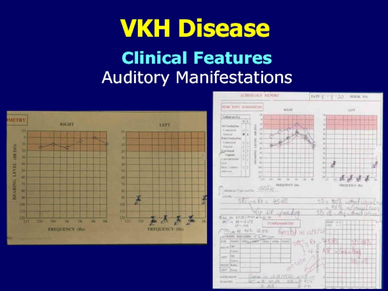
- 16. VKH DiseaseClinical Features Extraocular ManifestationsAuditory ManifestationsMay be
- 17. VKH DiseaseClinical Features Auditory Manifestations
- 18. VKH Disease 4 phases Prodromal Acute uveitic Convalescent or chronic Chronic recurrentClinical Course
- 19. VKH DiseaseProdromal Phase:Mimics viral illnessNeurologic and auditory
- 20. VKH DiseaseAcute Uveitic Phase:Bilateral in 70% of
- 21. VKH DiseaseAcute Uveitic Phase: (cont.).Yellow-white lesions at
- 22. VKH DiseaseAcute Uveitic Phase: (cont.)No inflammation of
- 23. VKH DiseaseAcute Uveitic Phase: (cont.).Shallow anterior chamberElevated IOPAcute angle closure glaucomaClinical Course
- 24. VKH DiseaseConvalescent Phase:Integumentary and uvea depigmentation.Perilimbal vitiligo
- 25. VKH DiseaseConvalescent Phase: (cont.)Multiple small yellow well-circumscribed
- 26. VKH DiseaseChronic Recurrent Phase:Acute episodes of granulomatous
- 27. VKH DiseaseChronic Recurrent Phase:Patients with recurrent VKH
- 28. VKH DiseaseFluorescein angiographyIndocyanine green angiographyUltrasonographyOptical coherence tomographyMultifocal
- 29. VKH DiseaseUltrasonography:Diffuse low to medium thickening of choroid.Overlying exudative RD.Laboratory Investigations
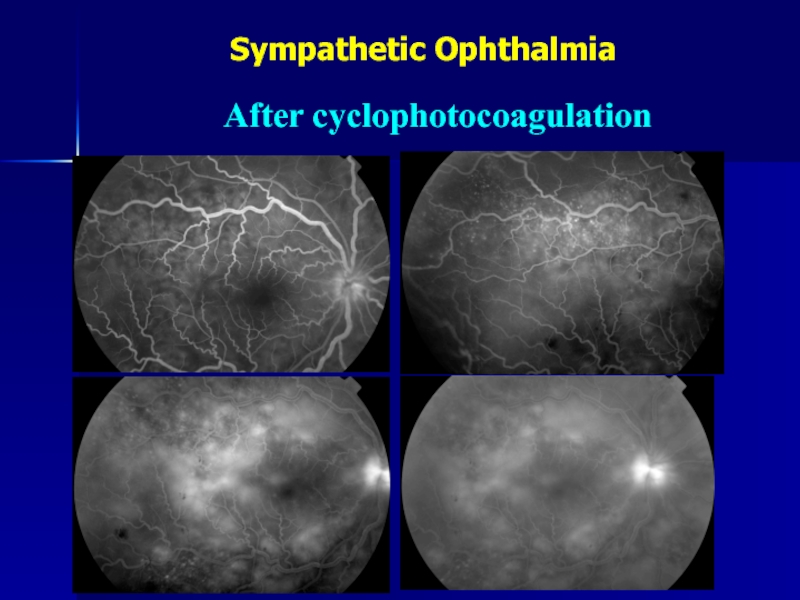
- 30. VKH DiseaseLaboratory InvestigationsOCTUseful in monitoring resolution of exudative retinal detachmentOn presentation4 weeks after systemic corticosteroid
- 31. VKH DiseaseMay be useful in detecting early
- 32. VKH DiseasePatients displayed a markedly decreased BCVA,
- 33. VKH DiseaseRetinal functional changes measured by microperimetry after immunosuppressive therapy
- 34. VKH DiseaseLumbar Puncture:Rarely necessary in a typical
- 35. VKH DiseaseLumbar Puncture:Frequency of CSF pleocytosis and
- 36. VKH DiseaseCataractSecondary glaucomaChoroidal neovascular membranesSubretinal fibrosisSevere chorioretinal
- 37. VKH DiseaseSignificantly associated with older age and
- 38. VKH DiseaseShould be prompt and aggressive.Systemic corticosteroids
- 39. VKH DiseaseIntravenous high-dose pulse steroid therapy (1g/day
- 40. VKH DiseaseSuch treatment may shorten duration of
- 41. VKH DiseaseFinal VA of 20/20 was significantly
- 42. VKH DiseasePatients treated initially with immunomodulatory drugs
- 43. VKH DiseaseUse of mycophenolate mofetil as first-line
- 44. VKH DiseaseVisual prognosis is generally favorable.87.5% achieved
- 45. VKH DiseasePoor visual acuity and severe anterior
- 46. Sympathetic OphthalmiaAhmed M. Abu El-Asrar, MD, PhD
- 47. Sympathetic OphthalmiaRare bilateral granulomatous panuveitis that occurs
- 48. Sympathetic OphthalmiaIn 0.1% to 0.3% of patients
- 49. Sympathetic OphthalmiaRole of ocular surgerySole cause in:45%
- 50. Sympathetic OphthalmiaRole of ocular surgeryOcular surgery, particularly
- 51. Sympathetic OphthalmiaRole of vitrectomyIn 0.06% of casesIn
- 52. Sympathetic OphthalmiaRole of vitrectomyVitreoretinal surgery is an
- 53. Sympathetic OphthalmiaEvisceration versus enucleationUveal tissue may be
- 54. Sympathetic OphthalmiaEvisceration versus enucleationFour cases of SO
- 55. Sympathetic OphthalmiaEvisceration versus enucleationEvisceration is safe with
- 56. Sympathetic OphthalmiaInterval between injury and onset of
- 57. Sympathetic Ophthalmia50-year-old man.Underwent successful retinal reattachment surgery
- 58. Sympathetic Ophthalmia31-year-old man.Underwent retinal reattachment surgery with
- 59. Sympathetic Ophthalmia50-year-old man.Developed SO 8 weeks after
- 60. Sympathetic Ophthalmia28-year-old man.Developed SO 12 weeks after
- 61. 38-year-old lady.Lt. eye blind since childhood with
- 62. Sympathetic OphthalmiaAfter cyclophotocoagulation
- 63. Sympathetic OphthalmiaVogt-Koyanagi-Harada disease(No previous ocular trauma or surgery)Differential Diagnosis
- 64. Sympathetic OphthalmiaImmunogeneticsGenetics predisposition which is very similar
- 65. Sympathetic OphthalmiaSimilar in both exciting and sympathizing
- 66. Sympathetic OphthalmiaPreventionCareful microsurgical management of the wound with prompt closure of all penetrating injuries.Management
- 67. Sympathetic OphthalmiaPreventionEnucleation of the traumatized eye if
- 68. Sympathetic OphthalmiaControversy regarding any advantage of enucleating
- 69. Sympathetic OphthalmiaEnucleation within 2 weeks of onset
- 70. Sympathetic OphthalmiaExciting eye may eventually have the
- 71. Sympathetic OphthalmiaIt is not justified to remove
- 72. Sympathetic OphthalmiaEarly diagnosis.Prompt and effective treatment with systemic immunosuppressive agents has improved the prognosis.Management
- 73. Sympathetic OphthalmiaCorticosteroids are the mainstay of treatment.I.V.
- 74. Sympathetic OphthalmiaSuccessful control of inflammation and
- 75. Sympathetic OphthalmiaRare disease.Major sight-threatening disorder.High index of
- 76. Sympathetic OphthalmiaDiagnosis is made clinically, histological proof
- 77. Sympathetic OphthalmiaPrognosis was considered poor prior to
- 78. Скачать презентанцию
Слайды и текст этой презентации
Слайд 1VOGT-KOYANAGI-HARADA
DISEASE
AHMED M. ABU EL-ASRAR, MD, PhD
Department of Ophthalmology, College
of Medicine, King Saud University, Riyadh, Saudi Arabia
Dr. Nasser Al-Rashid Research Chair in OphthalmologyСлайд 2Multisystem disease
Chronic, bilateral, granulomatous panuveitis associated with central nervous system,
auditory and integumentary manifestations
VKH Disease
Moorthy et al: Surv
Ophthalmol 1995; 39:265 (review)Read et al: Am J Ophthalmol 2001;131:647
Слайд 3VKH Disease
Individuals with a predisposing genetic background.
Ethnic groups with more
heavily pigmented skin.
Asians, Native Americans, Hispanics, Asian Indians, Middle Easterners.
Epidemiology
Слайд 4VKH Disease
Genetic background rather than degree of skin pigmentation.
Women more
than men.
3rd – 4th decade.
Pediatric age group.
Epidemiology
Read et al: Curr
Opin Ophthalmol 2000;11:437 (review)Abu El-Asrar et al: Eye 2008;22:1124
Martin et al: Retina 2010 Feb 17. [Epub ahead of print]
Слайд 5VKH Disease
Remains unknown.
T-lymphocyte mediated autoimmunity directed against one or more
antigens found on or associated with melanocytes found in eye,
skin and hair, inner ear, CNS.Etiology and Pathogenesis
Okada et al: Graefe’s Arch Clin Exp Ophthalmol 1996;234:359
Слайд 6VKH Disease
Tyrosinase family proteins are enzymes for melanin formation and
are expressed in melanocytes.
T-lymphocytes from VKH disease patients proliferate in
response to tyrosinase, TRP1 or TRP2.Immunization of Lewis rats with tyrosinase, TRP1 or TRP2 produced an inflammatory disease that resembled VKH disease with skin lesions and meningitis.
Yamaki et al: J Immunol 2000;165:7323
Yamaki et al: Exp Eye Res 2000;71:361
Etiology and Pathogenesis
Слайд 7VKH Disease
Th cells from peripheral blood of VKH patients produce
predominantly Th1 cytokines (IFN-gamma, IL-2) especially when stimulated
T-cell clones specific
to tyrosinase family proteins established from peripheral blood mononuclear cells of patients with VKH disease showed proliferative responses to tyrosinase and/or TRP1 and produced Th1-type cytokines.Imai et al: Curr Eye Res 2001;22:312
Gocho et al: Invest Ophthalmol Vis Sci 2001;42:2004
Etiology and Pathogenesis
Слайд 8VKH Disease
IL-23 stimulated production of IL-17 by CD4+
T cells may be responsible for the development of VKH
disease.Fang and Yang: Curr Eye Res 2008;33:517
Etiology and Pathogenesis
Слайд 9VKH Disease
VKH-like disease in patients treated with interferon-alpha and ribavirin
therapy for chronic hepatitis
C virus infection.Al-Muammar et al: Int Ophthalmol 2010 Feb 23. [Epub ahead of print]
Sene et al: World J Gastroenterol 2007;13:3137
Touitou et al: Am J Ophthalmol 2005;140:949
Papastathopoulos et al: J Infect 2006;52:e59
Kasahara et al: J Gastroenterol 2004;39:1106
Sylvestre et al: J Viral Hepat 2003;10:467
Etiology and Pathogenesis
Слайд 10VKH Disease
Granulomatous panuveitis.
Lymphocytes, epitheloid cells, few plasma cells, multinucleated giant
cells.
Epitheloid cells and giant cells contain melanin pigment.
Pathology
Слайд 11VKH Disease
Dalen-Fuchs’ nodules: Lymphocytes, epitheloid cells, pigment-laden macrophages, altered and/or
proliferated RPE cells.
Melanocytes disappear from choroid.
Perry and Font: Am J
Ophthalmol 1977;83:242Inomata and Rao: Am J Ophthalmol 2001;131:607
Pathology
Слайд 12VKH Disease
Certain racial groups.
Immunogenetic predisposition.
Strong association with HLA-DR4 and HLA-DRw53
with the most significant risk allele being HLA-DRB1*0405.
Causative pathogenic
antigen binds with HLA-DRB1*0405 molecule which presents the antigen to T cells to activate them.Fang and Wang: Curr Eye Res 2008;33:517 (review).
Read et al: Curr Opin Ophthalmol 2000;11:437 (review).
Yamaki et al: Int Ophthalmol Clin 2002;42:13 (review).
Genetic Factors
Слайд 13VKH Disease
VKH disease in monozygotic twins
Familial VKH disease
Familial cases shared
HLA-DR4
Itho et al: Int Ophthalmol 1992;16:49
Rutzen et al:
Am J Ophthalmol 1995;119:239Sonoda et al: Jpn J Ophthalmol 1999;43:113
Genetic Factors
Слайд 14VKH Disease
Integumentary Manifestations
Sensitivity of hair and skin to touch (early
in prodromal phase).
Poliosis, vitiligo, alopecia (during convalescent stage).
Ethnic groups may
manifest varying systemic symptoms. Clinical Features
Extraocular Manifestations
Слайд 15Clinical Features
Extraocular Manifestations
VKH Disease
Neurologic Manifestations
Most common during prodromal stage.
Neck stiffness,
headache, confusion.
Occasionally focal neurologic signs.
CSF pleocytosis.
Слайд 16VKH Disease
Clinical Features
Extraocular Manifestations
Auditory Manifestations
May be presenting problem
Sensorineural hearing loss
usually involves higher frequencies
Tinnitus
Vertigo
May cause permanent hearing loss
Слайд 18VKH Disease
4 phases
Prodromal
Acute uveitic
Convalescent or chronic
Chronic recurrent
Clinical Course
Слайд 19VKH Disease
Prodromal Phase:
Mimics viral illness
Neurologic and auditory manifestations
Few days
Headache, orbital
pain, stiff neck, malaise, abdominal pain, nausea, fever, vertigo, tinnitus
Cranial
nerve palsies, optic neuritis (rare)CSF pleocytosis
Clinical Course
Слайд 20VKH Disease
Acute Uveitic Phase:
Bilateral in 70% of patients, delay of
1-3 days before 2nd eye becomes involved in 30%. In
a few cases this interval may last up to 10 days.Hallmark is bilateral multifocal exudative retinal detachments, hyperemia and edema of the optic disc.
Clinical Course
Слайд 21VKH Disease
Acute Uveitic Phase: (cont.).
Yellow-white lesions at level of RPE
beneath serous RD.
Thickening of posterior choroid manifested by elevation of
peripapillary retino-choroid layer.Retinal edema in posterior pole.
Peripheral well-circumscribed yellow-white lesions (clinical equivalent of Dalen-Fuchs’ nodules).
Clinical Course
Слайд 22VKH Disease
Acute Uveitic Phase: (cont.)
No inflammation of the anterior segment
or mild to moderate nongranulomatous anterior uveitis if the disease
is not well controlled with appropriate treatment during the first two weeks.Clinical Course
Fang and Yang: Curr Eye Res 2008;33:517 (review)
Слайд 23VKH Disease
Acute Uveitic Phase: (cont.).
Shallow anterior chamber
Elevated IOP
Acute angle closure
glaucoma
Clinical Course
Слайд 24VKH Disease
Convalescent Phase:
Integumentary and uvea depigmentation.
Perilimbal vitiligo (Sugiura’s sign).
Fundus exhibits
an orange-red discoloration (“sunset-glow” fundus).
Clinical Course
Слайд 25VKH Disease
Convalescent Phase: (cont.)
Multiple small yellow well-circumscribed areas of chorioretinal
atrophy representing regressed Dalen-Fuchs’ nodules.
RPE clumping or migration.
Pigmented demarcation lines.
Clinical
Course
Слайд 26VKH Disease
Chronic Recurrent Phase:
Acute episodes of granulomatous anterior uveitis with
development of iris nodules
Recurrent posterior uveitis is distinctly uncommon
Complications are
seen in this phaseClinical Course
Слайд 27VKH Disease
Chronic Recurrent Phase:
Patients with recurrent VKH disease had a
more intensive inflammation in the anterior segment and long-lasting dysfunction
of the blood-aqueous barrier than those with initial onset VKH disease.Clinical Course
Fang et al: Br J Ophthalmol 2008;92:182
Слайд 28VKH Disease
Fluorescein angiography
Indocyanine green angiography
Ultrasonography
Optical coherence tomography
Multifocal electroretinograms
Lumbar puncture
Laboratory Investigations
Diagnosis
is made by clinical examination and ancillary test findings
Слайд 29VKH Disease
Ultrasonography:
Diffuse low to medium thickening of choroid.
Overlying exudative RD.
Laboratory
Investigations
Слайд 30VKH Disease
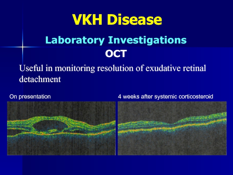
Laboratory Investigations
OCT
Useful in monitoring resolution of exudative retinal detachment
On
presentation
4 weeks after systemic corticosteroid
Слайд 31VKH Disease
May be useful in detecting early retinal damage.
Macular function
is severely impaired in patients with active uveitis.
Treatment with immunosuppressive
agents leads to delayed but limited recovery of macular function.May be useful in guiding therapy.
Laboratory Investigations
Chee et al: Graefe’s Arch Clin Exp Ophthalmol 2005; 243:785.
Multifocal Electroretinogram
Yang et al: Am J Ophthalmol 2008; 146:767.
Слайд 32VKH Disease
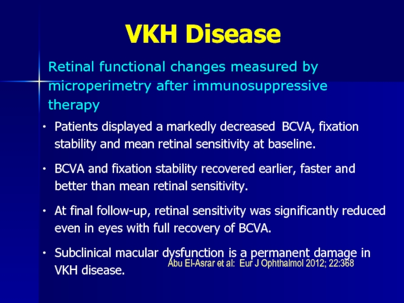
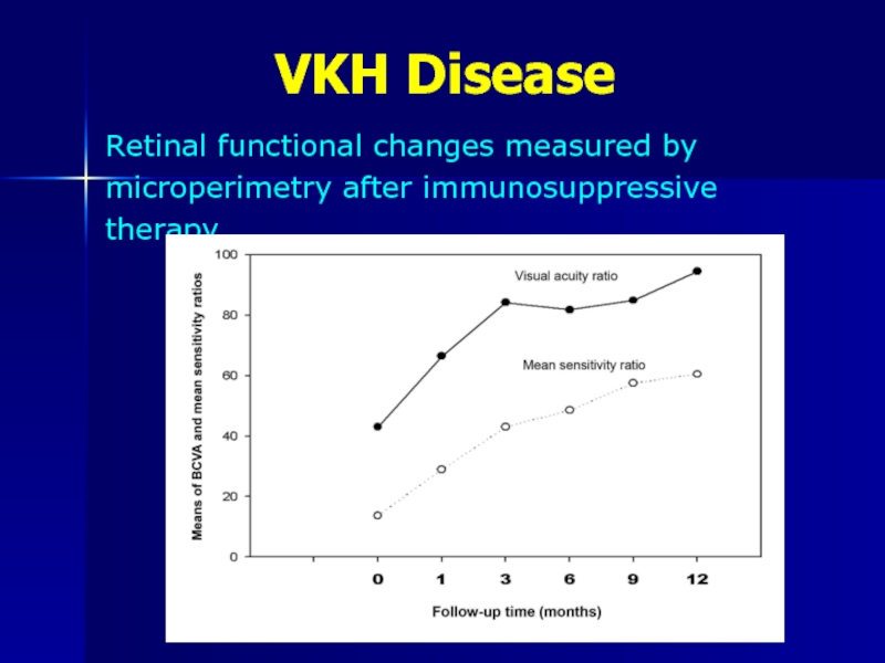
Patients displayed a markedly decreased BCVA, fixation stability and
mean retinal sensitivity at baseline.
BCVA and fixation stability recovered earlier,
faster and better than mean retinal sensitivity.At final follow-up, retinal sensitivity was significantly reduced even in eyes with full recovery of BCVA.
Subclinical macular dysfunction is a permanent damage in VKH disease.
Retinal functional changes measured by microperimetry after immunosuppressive therapy
Abu El-Asrar et al: Eur J Ophthalmol 2012; 22:368
Слайд 33VKH Disease
Retinal functional changes measured by microperimetry after immunosuppressive therapy
Слайд 34VKH Disease
Lumbar Puncture:
Rarely necessary in a typical case.
CSF pleocytosis (mostly
lymphocytes).
Transient and resolves within 8 weeks.
Laboratory Investigations
Слайд 35VKH Disease
Lumbar Puncture:
Frequency of CSF pleocytosis and the number of
cells in CSF at disease onset were significantly higher in
patients who eventually developed sunset glow fundus.Laboratory Investigations
Keino et al: Am J Ophthalmol 2006; 141:1140.
Слайд 36VKH Disease
Cataract
Secondary glaucoma
Choroidal neovascular membranes
Subretinal fibrosis
Severe chorioretinal atrophy
Significantly associated with
longer duration of disease and greater numbers of recurrences.
Complications
Read et
al : Am J Ophthalmol 2001;131:599.Sonoda et al: Jpn J Ophthalmol 1999;43:113.
Слайд 37VKH Disease
Significantly associated with older age and more severe disease
at presentation.
Complications
Al-Kharashi, Abu El-Asrar: Int Ophthalmol
2007;27:201Слайд 38VKH Disease
Should be prompt and aggressive.
Systemic corticosteroids are mainstay of
therapy.
1-1.5 mg/kg/day of oral Prednisone (single morning-after-breakfast dose).
For 6-12 months
with slow gradual tapering during this time.Hospitalization with careful follow-up.
Therapy
Слайд 39VKH Disease
Intravenous high-dose pulse steroid therapy (1g/day of Methylprednisolone given
for 3 days) followed by oral Prednisone (1 mg/kg/day).
Topical Prednisone
1% solution and cycloplegics for anterior uveitis.Patients adequately treated with corticosteroids have a fair visual prognosis.
Recurrences are associated with rapid or early decrease in steroid doses.
Therapy
Слайд 40VKH Disease
Such treatment may shorten duration of disease, prevent progression
into chronic stage, reduce incidence of extraocular manifestations.
Failure to prescribe
proper corticosteroid therapy in initial phase may lead to chronic recurrent uveitis that may result in severe visual loss due to extensive chorioretinal atrophy. Therapy
Sonoda et al: Jpn J Ophthalmol 1999;43:113.
Слайд 41VKH Disease
Final VA of 20/20 was significantly associated with use
of systemic corticosteroids for longer than 9 months and slow
tapering.Recurrent inflammation was significantly associated with rapid tapering of systemic corticosteroids.
Therapy
Al-Kharashi, Abu El-Asrar: Int Ophthalmol 2007;27:201
Слайд 42VKH Disease
Patients treated initially with immunomodulatory drugs (mycophenolate mofetil, cyclosporine
A, azathioprine, and methotrexate) combined with corticosteroids had a better
visual outcome than those who received corticosteroids as monotherapy.Immunomodulatory therapy combined with corticosteroids should be considered as first-line therapy for patients with VKH.
Therapy
Paredes et al: Ocul Immunol Inflamm 2006;14:87
Kim and Yu: Ocul Immunol Inflamm 2007;15:381
Abu El-Asrar, et al: Acta Ophthalmol 2012;90:e603
Слайд 43VKH Disease
Use of mycophenolate mofetil as first-line therapy combined with
systemic corticosteroids is safe and effective in the treatment of
acute uveitis associated with VKH disease.It has marked corticosteroid-sparing effect and significantly reduced development of chronic recurrent inflammation and late complications and significantly improved visual outcome.
The outcomes of mycophenolate mofetil combined with systemic corticosteroids in acute uveitis associated with VKH disease
Abu El-Asrar et al: Acta Ophthalmol 2012; 90:e603
Слайд 44VKH Disease
Visual prognosis is generally favorable.
87.5% achieved V.A. of ≥20/40.
High-dose
systemic corticosteroids for >9 months with slow tapering significantly improves
the prognosis and decreases risk of recurrence.Age older than 18 years is significantly associated with the development of complications.
Visual prognosis is generally favorable in children.
Prognosis
Al-Kharashi, Abu El-Asrar: Int Ophthalmol 2007;27:201
Abu El-Asrar et al: Eye 2008;22:1124
Слайд 45VKH Disease
Poor visual acuity and severe anterior segment inflammation at
presentation are significantly associated with a worse outcome.
Chronic recurrent disease
is significantly associated with more severe anterior segment inflammation and less exudative retinal detachment at presentation, more ocular complications and a worse visual outcome compared with initial-onset disease.Use of immunomodulatory therapy as first-line therapy combined with systemic corticosteroids significantly improved clinical outcomes.
Prognostic factors for clinical outcomes in patients treated with high-dose corticosteroids
Abu El-Asrar et al: Acta Ophthalmol. In press.
Слайд 47Sympathetic Ophthalmia
Rare bilateral granulomatous panuveitis that occurs as a complication
of a penetrating injury that involves the uvea of one
eye.Accidental trauma or surgery.
Injured eye is referred to as the exciting eye and fellow eye as the sympathizing eye.
Слайд 48Sympathetic Ophthalmia
In 0.1% to 0.3% of patients after accidental trauma.
In
0.015% of patients following ocular surgery.
5.8% and 0.67% after noncontact
and contact Nd:YAG cyclotherapy, respectively.Incidence
Allen: JAMA 1969;209:1090
Liddy, Stuart: Can J Ophthalmol 1972;7:157
Gass: Am J Ophthalmol 1982;93:552
Lam et al: Ophthalmology 1992;99:1818
Слайд 49Sympathetic Ophthalmia
Role of ocular surgery
Sole cause in:
45% of cases (Gass:
Am J Ophthalmol 1982;93:552)
17% of cases (Hakin et al: Eye
1992;6:453)28% of cases (Chan et al: Arch Ophthalmol 1995;113:597)
56% of cases (Kilmartin et al: Br J Ophthalmol 2000;84:259)
70% of cases (Su and Chee: Graefes’ Arch Clin Exp Ophthalmol
2006;244:243)
38% of cases (Galor et al: Am J Ophthalmol 2009;148:704)
Incidence
Слайд 50Sympathetic Ophthalmia
Role of ocular surgery
Ocular surgery, particularly retinal surgery, is
now a greater risk than accidental trauma.
Risk of one in
1152 retinal surgical procedures.Incidence
Kilmartin et al: Br J Ophthalmol 2000;84:448
Kilmartin et al: Br J Ophthalmol 2000;84:259
Su and Chee: Graefe’s Arch Clin Exp Ophthalmol 2006;244:243
Слайд 51Sympathetic Ophthalmia
Role of vitrectomy
In 0.06% of cases
In 0.01% when vitrectomy
was the only operative procedure and penetrating wound.
A prospective surveillance
for SO in UK and Ireland showed risk of one in 799 vitrectomies.Incidence
Kilmartin et al: Br J Ophthalmol 2000;84:448
Gass: Am J Ophthalmol 1982;93:552
Слайд 52Sympathetic Ophthalmia
Role of vitrectomy
Vitreoretinal surgery is an important risk factor.
It
may be appropriate to counsel patients about risk of SO
before performing vitrectomyAny bilateral uveitis following vitreoretinal surgery should alert the surgeon to the possibility of SO.
Incidence
Kilmartin et al: Br J Ophthalmol 2000;84:448
Слайд 53Sympathetic Ophthalmia
Evisceration versus enucleation
Uveal tissue may be left behind after
evisceration and act as the source of the immune response.
Controversy
involving evisceration and risk of SO.Incidence
Слайд 54Sympathetic Ophthalmia
Evisceration versus enucleation
Four cases of SO following evisceration
Six cases
of SO after evisceration.
A case of SO following evisceration of
a blind, painful, post-traumatic, glaucomatous eye.Incidence
Green et al: Trans Am Acad Ophthalmol Otolaryngol 1972; 76:625
Ikui et al: Nippon Ganka Kiyo1965; 16:458
Griepentrog et al: Ophthal Plast Reconstr Surg 2005; 21:316
Слайд 55Sympathetic Ophthalmia
Evisceration versus enucleation
Evisceration is safe with little risk of
SO.
High degree of clinical suspicion is required.
Incidence
Levine et
al: Ophthal Plast Reconstr Surg 1999; 15:4.Toit et al: Br J Ophthalmol 2008;92:61.
Слайд 56Sympathetic Ophthalmia
Interval between injury and onset of inflammation ranges from
5 days to 66 years.
70 to 80% occur within 3
months and 90% within 1 year of injury.Peak incidence occurs at 4 to 8 weeks following injury.
Clinical Manifestations
Lubin et al: Ophthalmology 1980;87:109
Zaharia et al: Can J Ophthalmol 1984;19:240
Manak: Surv Ophthalmol 1979;24:141
Слайд 57Sympathetic Ophthalmia
50-year-old man.
Underwent successful retinal reattachment surgery with pars plana
vitrectomy and gas tamponade.
Developed SO 5 weeks after surgery
Treatment: I.V.
and oral steroids + cyclosporin A.Control of inflammation and good visual prognosis.
After successful retinal reattachment surgery with vitrectomy
Слайд 58Sympathetic Ophthalmia
31-year-old man.
Underwent retinal reattachment surgery with silicone oil tamponade
in his left eye 3 mos. prior to presentation.
VA: CF
OUTreatment: I.V. and oral steroids + mycophenolate mofetil
Control of inflammation and good visual prognosis.
After retinal reattachment surgery with silicone oil tamponade
Слайд 59Sympathetic Ophthalmia
50-year-old man.
Developed SO 8 weeks after cataract surgery.
Treatment: Oral
steroids + cyclosporin A + pars plana vitrectomy and removal
of IOL.Control of inflammation and good visual prognosis.
After complicated cataract surgery and IOL implantation
Слайд 60Sympathetic Ophthalmia
28-year-old man.
Developed SO 12 weeks after sustaining penetrating trauma.
Treatment:
Oral steroids + cyclosporin A.
Control of inflammation and good visual
outcome.After penetrating trauma
Слайд 6138-year-old lady.
Lt. eye blind since childhood with no known clear
cause.
Developed SO 9 weeks after left eye cyclophotocoagulation that was
done prior to referral.Sympathetic Ophthalmia
After cyclophotocoagulation
Слайд 63Sympathetic Ophthalmia
Vogt-Koyanagi-Harada disease
(No previous ocular trauma or surgery)
Differential Diagnosis
Слайд 64Sympathetic Ophthalmia
Immunogenetics
Genetics predisposition which is very similar to VKH disease.
Increased
frequency of HLA-A11 antigen in patients with SO.
Associated with HLA-DRB1*04,
DQA1*03, DQB1*04 among Japanese, British, Irish patients.Pathogenesis
Reynard et al: Am J Ophthalmol 1983;95:216
Shindo et al; Tissue Antigens 1997;49:111
Kilmartin et al: Br J Ophthalmol 2001;85:281.
Слайд 65Sympathetic Ophthalmia
Similar in both exciting and sympathizing eyes.
Classic description is
that of a diffuse non-necrotizing granulomatous choroidal inflammation with Dalen-Fuchs
nodules.Relative sparing of the choriocapillaris or retina (uncharacteristic feature).
Pathological Features
Слайд 66Sympathetic Ophthalmia
Prevention
Careful microsurgical management of the wound with prompt closure
of all penetrating injuries.
Management
Слайд 67Sympathetic Ophthalmia
Prevention
Enucleation of the traumatized eye if unsalvageable by modern
surgical methods within two weeks after injury is the only
known preventive way.Problems:
No proof that this is actually of value.
Incidence of SO after penetrating injury is decreasing.
With current advanced surgical techniques many eyes may have a fair prognosis.
Management
Слайд 68Sympathetic Ophthalmia
Controversy regarding any advantage of enucleating the exciting eye
once SO has started in the sympathizing eye.
Management
Слайд 69Sympathetic Ophthalmia
Enucleation within 2 weeks of onset is associated with
a relatively benign clinical course and improves visual outcome.
Enucleation is
valueless and should not be performed.Enucleation of the exciting eye did not result in improved visual function in the sympathizing eye.
Management
Winter: Am J Ophthalmol 1955;39:340
Lubin et al: Ophthalmology 1980;87:109
Reynard et al: Am J Ophthalmol 1983;96:290
Слайд 70Sympathetic Ophthalmia
Exciting eye may eventually have the better vision, or
diagnosis may be incorrect.
Enucleation should be considered only in eyes
with nil visual prognosis.Management
Слайд 71Sympathetic Ophthalmia
It is not justified to remove a functionally useful
injured eye in established cases of SO for the purpose
of decreasing inflammation.Not all inciting eyes are “lost eyes” as commonly believed.
Management
Слайд 72Sympathetic Ophthalmia
Early diagnosis.
Prompt and effective treatment with systemic immunosuppressive agents
has improved the prognosis.
Management
Слайд 73Sympathetic Ophthalmia
Corticosteroids are the mainstay of treatment.
I.V. pulses, I g/day
methylprednisolone, 3 consecutive days for immediate control
of inflammation.Followed by oral prednisone 1-1.5 mg/ Kg/day.
Dose is reduced gradually following clinical resolution of uveitis.
Continued for at least 6 months.
Management
Слайд 74Sympathetic Ophthalmia
Successful control of inflammation and good visual prognosis
is related to prompt and adequate systemic immunosuppression using combination
of systemic steroids and steroid sparing agents such as cyclosporin A, azathioprine, mycophenolate mofetil.Management
- Hakin et al: Eye 1992;6:453
Chang et al: Arch Ophthalmol 1995;113:597
Kilmartin et al: Br J Ophthalmol 2000;84:259
Abu El-Asrar, Al-Obeidan: Eur J Ophthalmol 2001;11:193
Слайд 75Sympathetic Ophthalmia
Rare disease.
Major sight-threatening disorder.
High index of suspicion must be
maintained whenever inflammation occurs in fellow eyes of an eye
that has suffered penetrating trauma or intraocular surgery.Infection should be carefully ruled out.
Conclusions
Слайд 76Sympathetic Ophthalmia
Diagnosis is made clinically, histological proof is not required.
Injured
eyes which have potential vision should not be enucleated in
an attempt to prevent or lessen SO or to provide confirmatory pathology.Conclusions
Слайд 77Sympathetic Ophthalmia
Prognosis was considered poor prior to the use of
systemic immunosuppression.
Today, it should no longer be regarded as a
blinding disease.Prompt and adequate systemic immunosuppressive therapy with systemic steroids and steroid-sparing agents has improved prognosis.
Conclusions

