Слайд 1Assessment of respiratory system
Dr .Essmat Gemaey
Assistant prof.Psychiatric nursing
Слайд 2Learning objectives
After completion of this session the students should be
able to:
Revise knowledge of anatomy and physiology
Obtain health history
about respiratory system
Demonstrate physical examination
Differentiate between normal and abnormal findings
Слайд 3Outlines
anatomy and physiology of respiratory system
Assessment of respiratory system
]
1
Position/Lighting/Draping
2 Inspection
2.1 Chest wall deformities
2.2 Signs of
respiratory distress
3 Palpation
4 Percussion
5 Ausculation
5.1 Vocal fremitus (not usually done)
Слайд 4Anatomy and physiology
The respiratory tract extends from the nose
to the alveoli and includes not only the air-conducting passages
also but the blood supply
The primary purpose of the respiratory system is gas exchange, which involves the transfer of oxygen and carbon dioxide between the atmosphere and the blood.
The respiratory system is divided into two parts: the upper respiratory tract and the
lower respiratory tract
Слайд 5
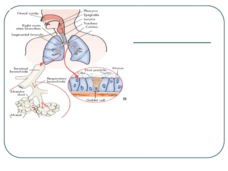
The nose
pharynx
adenoids
tonsils
epiglottis
larynx,
and trachea.
The
upper respiratory tract includes
Слайд 6The lower respiratory tract consists of
the bronchi,
Bronchioles
alveolar ducts
and
alveoli
With the exception of the right and left main-stem
bronchi, all lower airway structures are contained within the lungs.
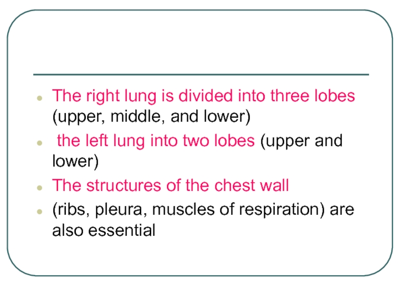
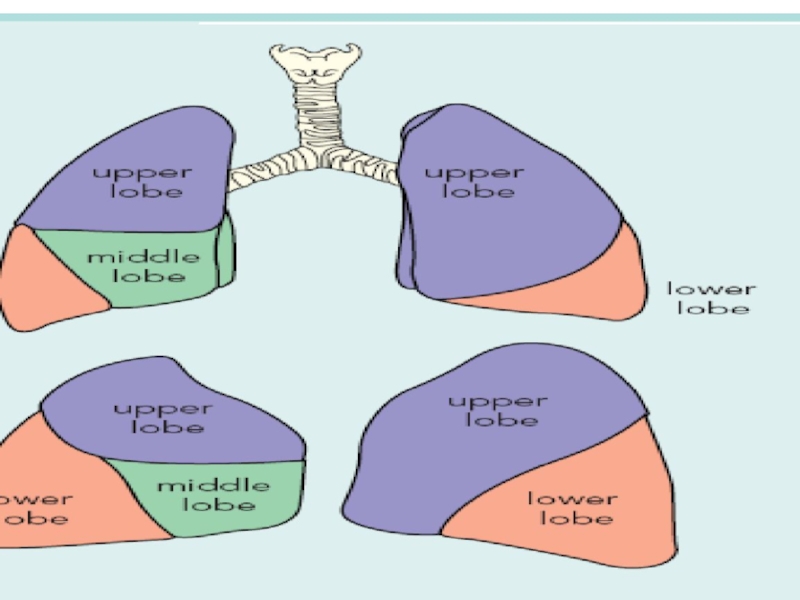
Слайд 7The right lung is divided into three lobes (upper, middle,
and lower)
the left lung into two lobes (upper
and lower)
The structures of the chest wall
(ribs, pleura, muscles of respiration) are also essential
Слайд 10Physiology of Respiration
Ventilation. Ventilation involves inspiration (movement of
air into the
lungs) and expiration (movement of air out of the
lungs). Air
moves in and out of the lungs because intrathoracic
pressure changes in relation to pressure at the airway opening.
Contraction of the diaphragm and intercostal and scalene muscles
increases chest dimensions, thereby decreasing intrathoracic
pressure. Gas flows from an area of higher pressure (atmospheric)
to one of lower pressure (intrathoracic)
Слайд 11Equipment Needed
A Stethoscope
A Peak Flow Meter
Слайд 12Surface markings of the lobes of the lung:
(a) anterior, (b)
posterior, (c) right lateral and (d) left lateral.
(UL, upper lobe;
ML, middle lobe; LL, lower lobe).
Ul
ml
a
b
ll
ul
ll
ul
ll
ml
Слайд 15Position/Lighting/Draping
Position –
patient should sit upright on the examination table.
The patient's hands should remain at their sides.
When the
back is examined the patient is usually asked to move their arms forward (hug themself position) so that the scapulae are not in the way of examining the upper lung fields.
Lighting - adjusted so that it is ideal.
Draping - the chest should be fully exposed. Exposure time should be minimized.
Слайд 16The basic steps of the examination
can be remembered with the
mnemonic IPPA:
Inspection
Palpation
Percussion
Auscultation
Слайд 17Health History
Any risk factors for respiratory disease
smoking
pack years
ppd X # years
exposure to smoke
history of attempts
to quit, methods, results
sedentary lifestyle, immobilization
age
environmental exposure
Dust, chemicals, asbestos, air pollution
obesity
family history
Слайд 18Cough
Type
dry, moist, wet, productive, hoarse, hacking, barking, whooping
Onset
Duration
Pattern
activities, time of day, weather
Severity
effect on ADLs
Wheezing
Associated
symptoms
Treatment and effectiveness
Слайд 19sputum
amount
color
presence of blood (hemoptysis)
odor
consistency
pattern of
production
Слайд 20Respiratory infections or diseases (URI)
Trauma
Surgery
Chronic conditions of
other systems
Family Health History
Tuberculosis
Emphysema
Lung Cancer
Allergies
Asthma
Past Health History
Слайд 21Inspection
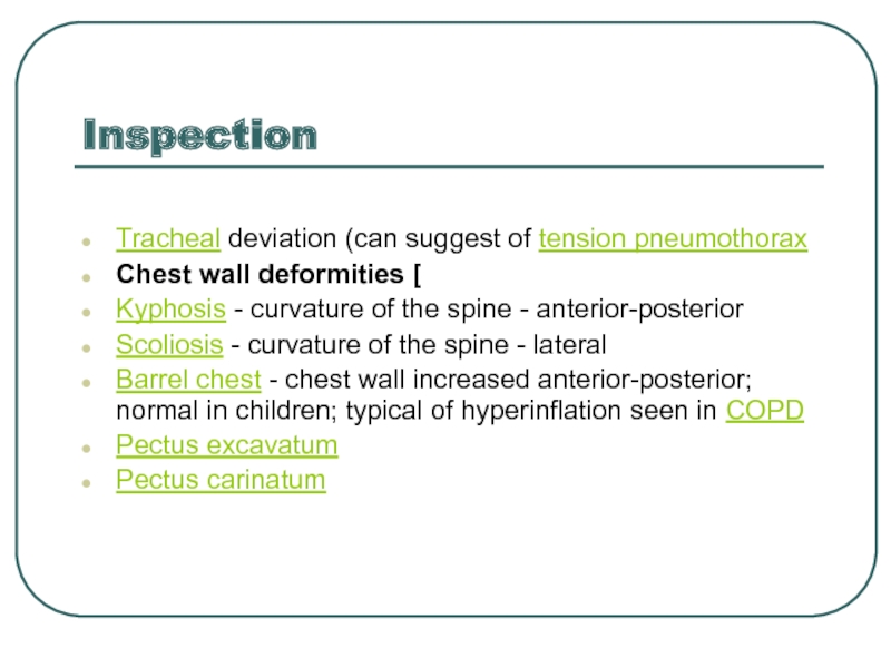
Tracheal deviation (can suggest of tension pneumothorax
Chest wall deformities [
Kyphosis
- curvature of the spine - anterior-posterior
Scoliosis - curvature
of the spine - lateral
Barrel chest - chest wall increased anterior-posterior; normal in children; typical of hyperinflation seen in COPD
Pectus excavatum
Pectus carinatum
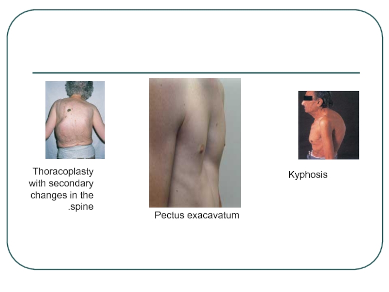
Слайд 22Kyphosis
Thoracoplasty
with secondary
changes in the
spine.
Pectus exacavatum
Слайд 23Signs of respiratory distress
Cyanosis - person turns blue
Pursed-lip breathing
- seen in COPD (used to increase end expiratory pressure)
Accessory muscle use (scalene muscles)
Diaphragmatic paradox - the diaphragm moves opposite of the normal direction on inspiration; suspect flail segment in trauma
Intercostal indrawing
Слайд 24‘pink puffer’. Note the
pursed-lip
breathing
.
‘blue bloater’
showing ascites
from marked cor
pulmonale.
Слайд 26Palpation
Tactile fremitus
is vibration felt by palpation. Place your open
palms against the upper portion of the anterior chest, making
sure that the fingers do not touch the chest. Ask the patient to repeat the phrase “ninety-nine” or another resonant phrase while you systematically move your palms over the chest from the central airways to each lung’s periphery.You should feel vibration of equally intensity on both sides of the chest. Examine the posterior thorax in a similar manner. The fremitus should be felt more strongly in the upper chest with little or no fremitus being felt in the lower chest
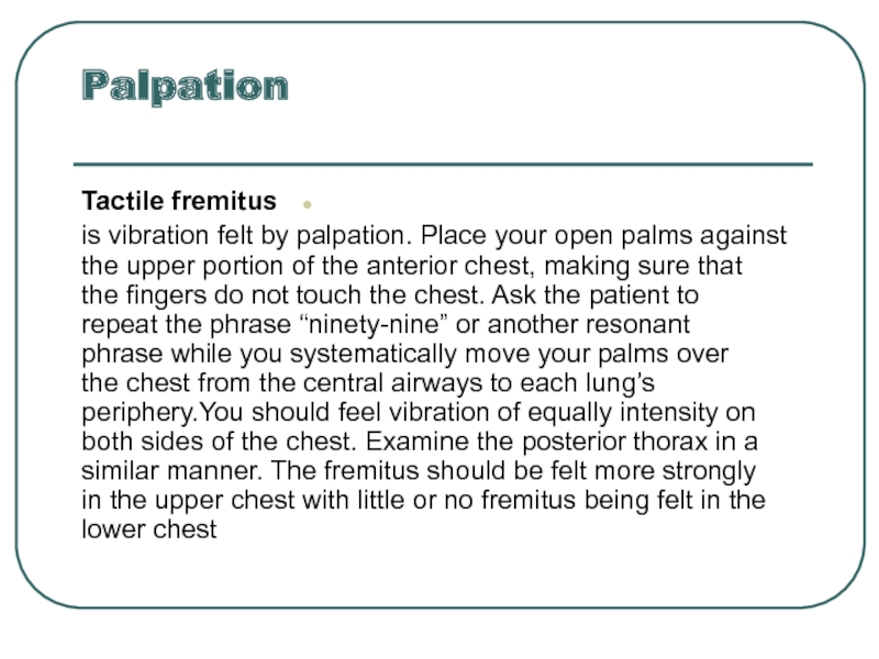
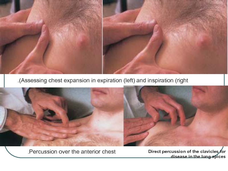
Слайд 27Assessing chest expansion in expiration (left) and inspiration (right).
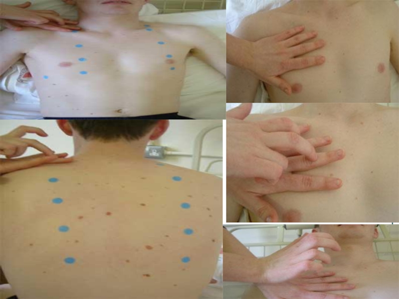
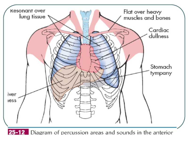
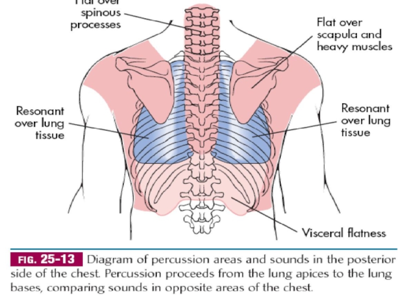
Direct percussion
of the clavicles for disease in the lung apices
Percussion over
the anterior chest.
Слайд 29Auscultation
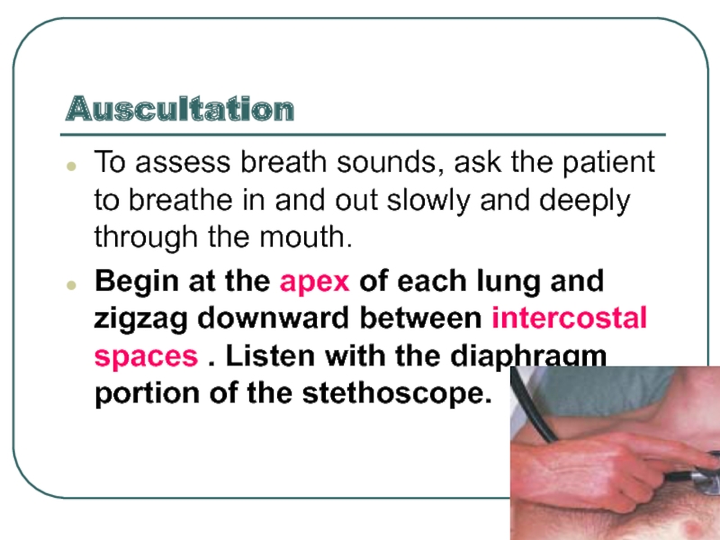
To assess breath sounds, ask the patient to breathe in
and out slowly and deeply through the mouth.
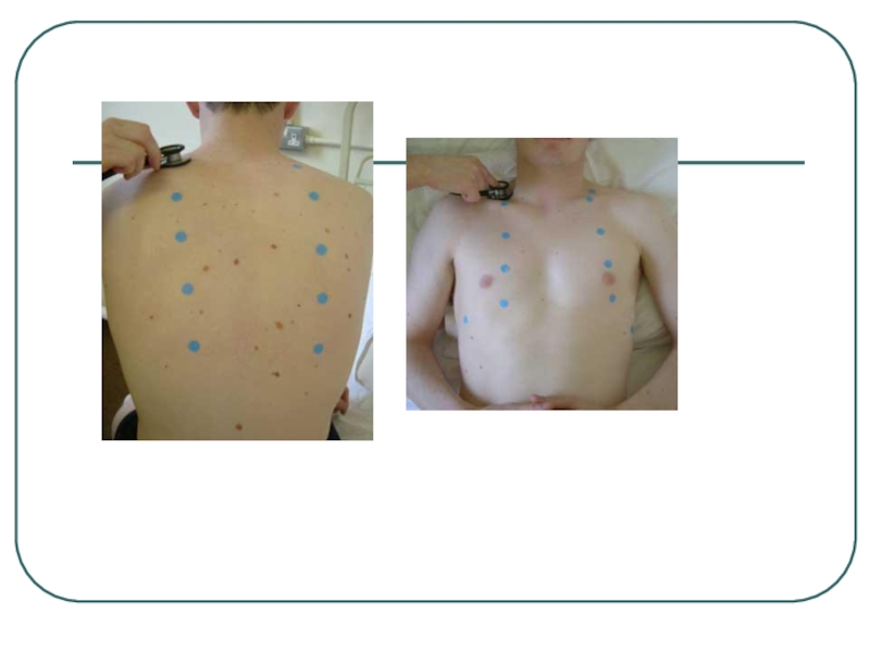
Begin at
the apex of each lung and zigzag downward between intercostal spaces . Listen with the diaphragm portion of the stethoscope.
Слайд 30Normal breath sounds
Note
Pitch
Intensity
Quality
Duration
Слайд 32Normal Breath Sounds
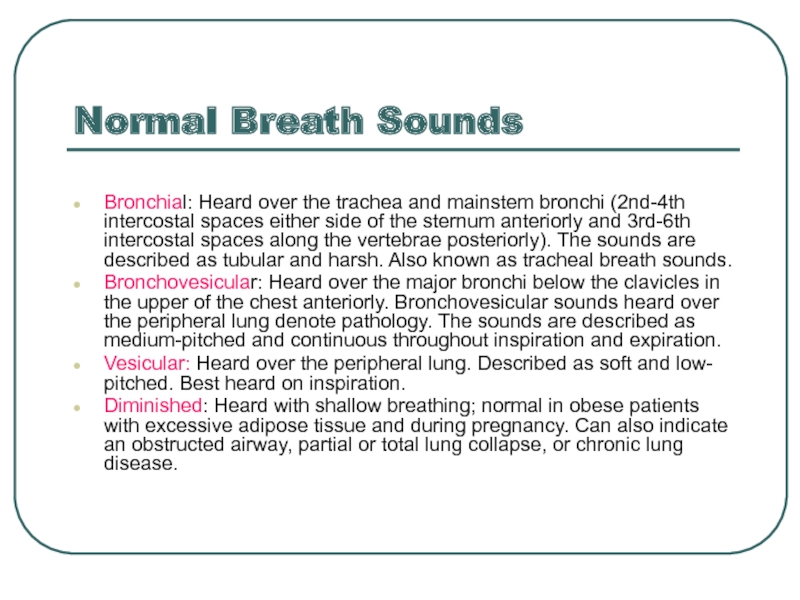
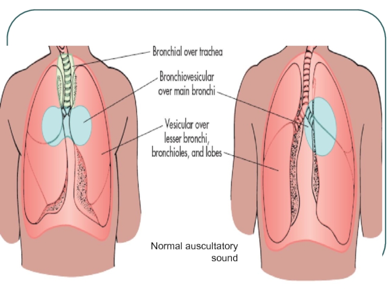
Bronchial: Heard over the trachea and mainstem bronchi
(2nd-4th intercostal spaces either side of the sternum anteriorly and
3rd-6th intercostal spaces along the vertebrae posteriorly). The sounds are described as tubular and harsh. Also known as tracheal breath sounds.
Bronchovesicular: Heard over the major bronchi below the clavicles in the upper of the chest anteriorly. Bronchovesicular sounds heard over the peripheral lung denote pathology. The sounds are described as medium-pitched and continuous throughout inspiration and expiration.
Vesicular: Heard over the peripheral lung. Described as soft and low- pitched. Best heard on inspiration.
Diminished: Heard with shallow breathing; normal in obese patients with excessive adipose tissue and during pregnancy. Can also indicate an obstructed airway, partial or total lung collapse, or chronic lung disease.
Слайд 35Tactile Fremitus
Ask the patient to say "ninety-nine" several times in
a normal voice.
Palpate using the ball of your hand.
You should feel the vibrations transmitted through the airways to the lung.
Increased tactile fremitus suggests consolidation of the underlying lung tissues

![Assessment of respiratory system Outlinesanatomy and physiology of respiratory system Assessment of respiratory system]1 Position/Lighting/Draping Outlinesanatomy and physiology of respiratory system Assessment of respiratory system]1 Position/Lighting/Draping 2 Inspection 2.1 Chest wall deformities](/img/thumbs/1293adadb1ca18d3a0ef3b1aa6a13939-800x.jpg)
