Разделы презентаций
- Разное
- Английский язык
- Астрономия
- Алгебра
- Биология
- География
- Геометрия
- Детские презентации
- Информатика
- История
- Литература
- Математика
- Медицина
- Менеджмент
- Музыка
- МХК
- Немецкий язык
- ОБЖ
- Обществознание
- Окружающий мир
- Педагогика
- Русский язык
- Технология
- Физика
- Философия
- Химия
- Шаблоны, картинки для презентаций
- Экология
- Экономика
- Юриспруденция
Chapter 21 Lecture Outline See separate PowerPoint slides for all figures and
Содержание
- 1. Chapter 21 Lecture Outline See separate PowerPoint slides for all figures and
- 2. 21.1 Lymph and Lymph VesselsDescribe
- 3. 21.1 Lymph and Lymph VesselsLymphatic system functionsTransport
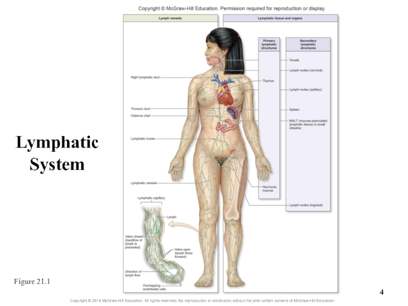
- 4. Lymphatic System Figure 21.1
- 5. TonsilsPalatinePharyngealLingual
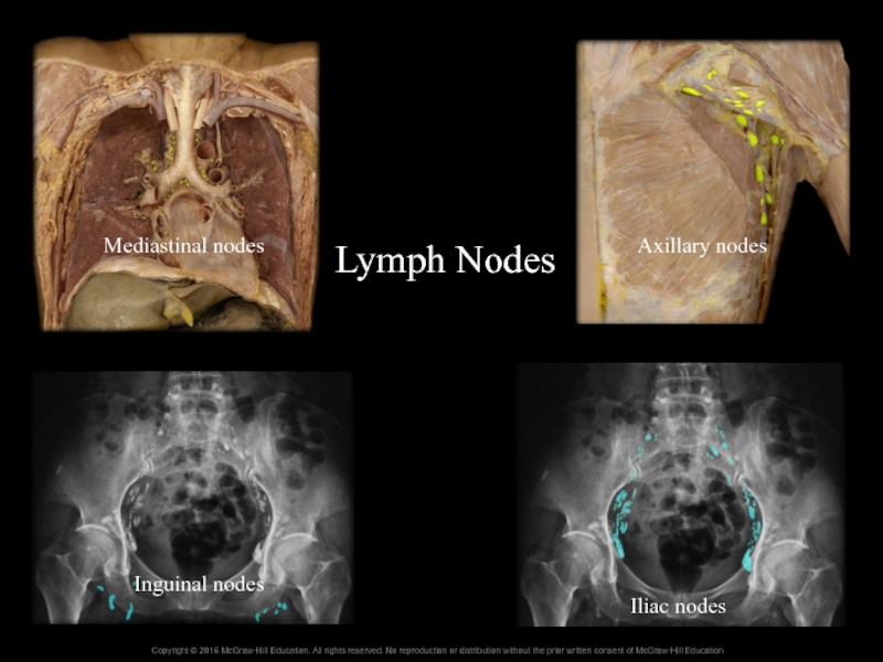
- 6. Lymph NodesMediastinal nodesAxillary nodesInguinal nodesIliac nodes
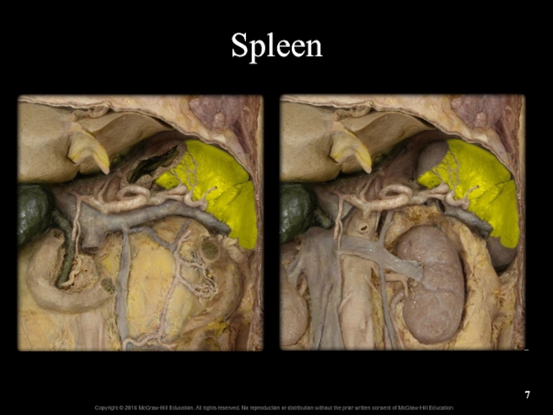
- 7. Spleen
- 8. 21.1a Lymph and Lymphatic CapillariesCharacteristics of lymphSome
- 9. 21.1a Lymph and Lymphatic CapillariesLymphatic capillariesSmall, closed-ended
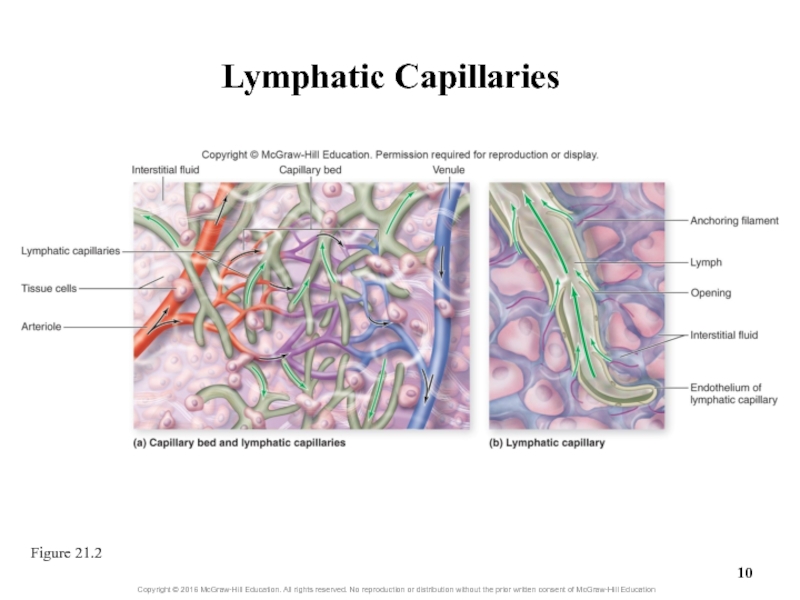
- 10. Lymphatic Capillaries Figure 21.2
- 11. 21.1a Lymph and Lymphatic CapillariesMovement of lymph
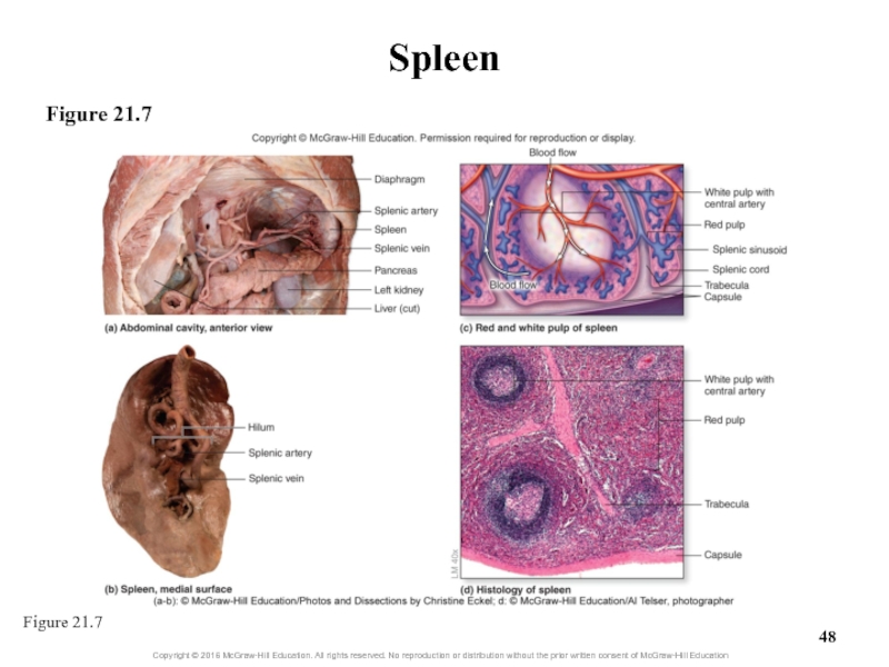
- 12. Clinical View: MetastasisWandering cancerous cells establishing secondary
- 13. Lymphatic Drainage of Mammary and Axillary
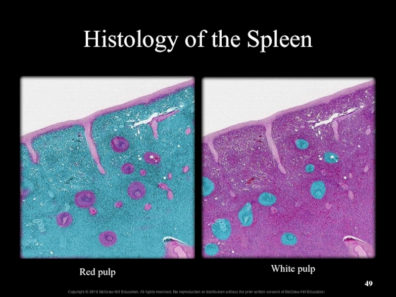
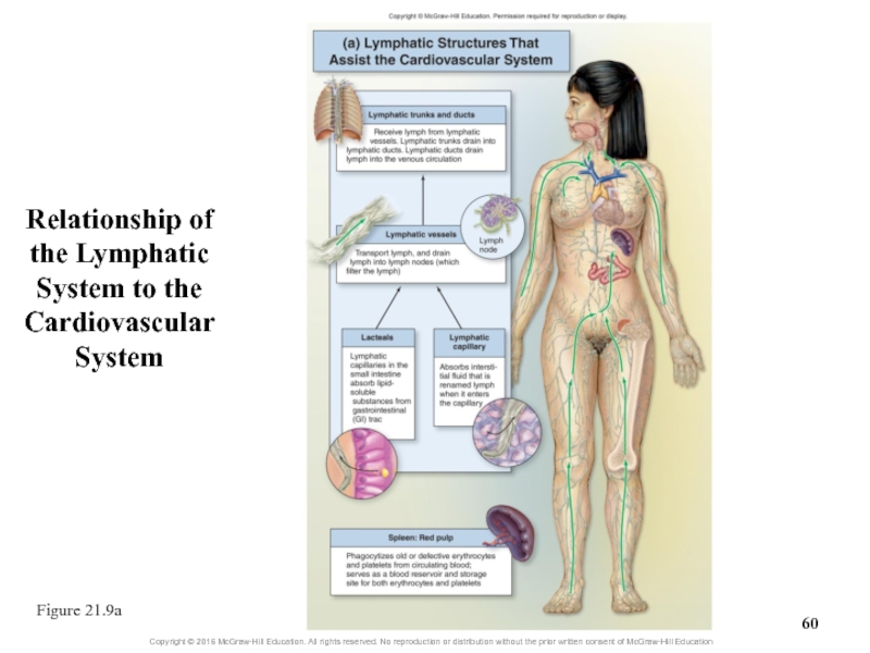
- 14. 21.1b Lymphatic Vessels, Trunks, and DuctsLymphatic vessels
- 15. 21.1b Lymphatic Vessels, Trunks, and DuctsLymphatic trunks
- 16. 21.1b Lymphatic Vessels, Trunks, and DuctsLymphatic ducts
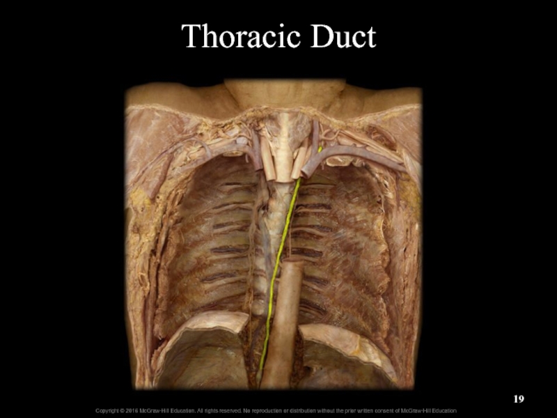
- 17. Lymphatic ducts (continued)Thoracic duct is largest lymphatic
- 18. Lymphatic Trunks and DuctsFigure 21.3a
- 19. Thoracic Duct
- 20. Cysterna Chyli
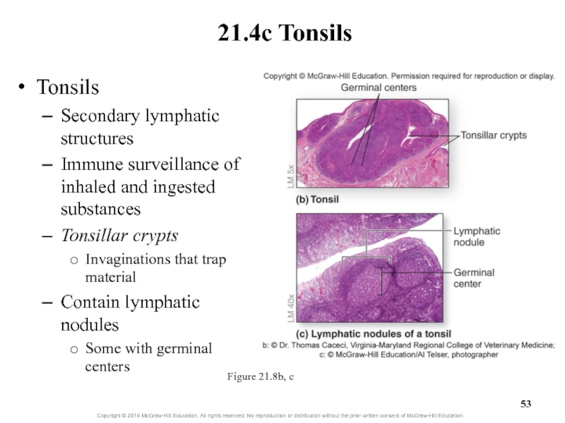
- 21. Clinical View: LymphedemaAccumulation of interstitial fluid due
- 22. What did you learn?How does fluid enter
- 23. 21.2 Overview of Lymphatic Tissue
- 24. 21.2 Overview of Lymphatic Tissue and OrgansPrimary
- 25. What did you learn?What does the acronym
- 26. Thymus
- 27. 21.3 Overview of Lymphatic Tissue
- 28. 21.3a Red Bone MarrowRed bone marrowLocated between
- 29. 21.3b ThymusThymus involved in T-lymphocyte maturationLocated in
- 30. Thymus GlandThymus (adult)Thymus (infant)
- 31. Figure 21.5aThymus
- 32. Figure 21.5b-cThymus
- 33. Fetal Thymus LM: Low MagnificationLobule of thymusLobe of thymusSepta of thymusCortex of thymusMedulla of thymus
- 34. What did you learn?Where does hemopoiesis occur?What is the function of the thymus?
- 35. 21.4 Secondary Lymphatic StructuresDescribe the
- 36. 21.4 Secondary Lymphatic StructuresSecondary lymphatic structuresHouse lymphocytes
- 37. 21.4a Lymph NodesLymph nodes filter lymph, remove
- 38. 21.4a Lymph NodesLymph node componentsAfferent lymphatic vessels
- 39. Node’s cortex contains lymphatic nodulesReticular fibers support
- 40. 21.4a Lymph NodesNode’s medulla contains medullary cordsConnective
- 41. Lymph Node HistologyCortexMedullaGerminal centersLymphadenitisLymphadenopathy
- 42. 21.4a Lymph NodesLymph flow through nodesLymph enters
- 43. Clinical View: LymphomaMalignant neoplasm from lymphatic structuresNontender,
- 44. 21.4b SpleenThe spleen is the largest lymphatic
- 45. Spleen
- 46. 21.4b SpleenTissues of the spleenWhite pulpClusters of
- 47. SpleenSplenic a.Splenic v.
- 48. SpleenFigure 21.7Figure 21.7
- 49. Histology of the SpleenRed pulpWhite pulp
- 50. Spleen LM: Medium MagnificationCapsuleRed pulpWhite pulpTrabeculaeGerminal center of white pulpCentral white pulp arteries
- 51. 21.4b SpleenMonitoring blood as it flows through
- 52. Clinical View: SplenectomySurgical removal of the spleenMay
- 53. TonsilsSecondary lymphatic structures Immune surveillance of inhaled
- 54. Tonsils (continued)Pharyngeal tonsilIn nasopharynxCalled adenoids when enlarged
- 55. TonsilsPalatinePharyngealLingual
- 56. Palatine Tonsil HistologyTonsilEpitheliumGerminal centersLymphatic nodulesTonsillar crypts
- 57. Clinical View: Tonsillitis and TonsillectomyAcute tonsillitis: inflammation
- 58. 21.4d Lymphatic Nodules and MALTLymphatic nodulesClusters of
- 59. 21.4d Lymphatic Nodules and MALTMALTMucosa-associated lymphatic tissueLocated
- 60. Relationship of the Lymphatic System to the Cardiovascular SystemFigure 21.9a
- 61. Relationship of the Lymphatic System to the Immune SystemFigure 21.9b
- 62. What did you learn?What is the typical
- 63. Скачать презентанцию
Слайды и текст этой презентации
Слайд 1Chapter 21
Lecture Outline
See separate PowerPoint slides for all figures and
tables pre-inserted into PowerPoint without notes.
Слайд 2
21.1
Lymph and Lymph Vessels
Describe lymph and its contents.
Discuss the
location and anatomic structure of lymphatic capillaries.
Explain how fluid enters
lymphatic capillaries.Explain the mechanisms that move lymph through lymphatic vessels, trunks, and ducts.
Name the five types of lymphatic trunks and the regions of the body from which they drain lymph.
Describe the regions that are drained by the right lymphatic duct and by the thoracic duct.
Learning Objectives:
Слайд 321.1 Lymph and Lymph Vessels
Lymphatic system functions
Transport and house lymphocytes
and other immune cells
Return excess fluid in body tissues to
blood to maintain blood volumeLymphatic system components
Lymph vessels, lymphatic issues and organs
Lymph is the fluid transported within lymph vessels
Слайд 821.1a Lymph and Lymphatic Capillaries
Characteristics of lymph
Some fluid leaves blood
capillaries and is not reabsorbed by them
Such fluid moves into
lymphatic capillaries where it is called lymphComponents of lymph
Water, dissolved solutes, and small amount of protein
Sometimes cell debris, pathogens, or cancer cells
Слайд 921.1a Lymph and Lymphatic Capillaries
Lymphatic capillaries
Small, closed-ended vessels that absorb
interstitial fluid
Interspersed around most blood capillaries
Absent in avascular tissues, red
marrow, spleen, and CNSSlightly larger than blood capillaries; no basement membrane
Walls are made of overlapping endothelial cells
Have flaps between cells through which fluid enters but can’t exit
Anchoring filaments hold endothelial cells to nearby structures
Lacteals: lymphatic capillaries in GI tract
Absorb lipid-soluble substances from GI tract
Слайд 1121.1a Lymph and Lymphatic Capillaries
Movement of lymph into lymphatic capillaries
Hydrostatic
pressure of interstitial fluid pushes it into capillary
Anchoring filaments linking
endothelial cells to surrounding structures prevent vessel collapsePressure of lymph inside vessel forces intercellular openings (“flaps”) of capillary wall to close with lymph inside
Lymph moves through vessels of larger and larger size
Lymphatic capillaries, lymphatic vessels, lymphatic trunks, and lymphatic ducts
Ultimately, fluid is returned to blood circulation
Слайд 12Clinical View: Metastasis
Wandering cancerous cells establishing secondary tumors
Develop in other
locations in the body (metastasis)
E.g., breast cancer may metastasize to
the lungCancerous cells break free from primary tumor
Transported in the lymph
Слайд 13Lymphatic Drainage of Mammary
and Axillary Regions
Axillary lymph nodes
Pectoralis major muscle
Слайд 1421.1b Lymphatic Vessels, Trunks, and Ducts
Lymphatic vessels are fed by
lymphatic capillaries
Located adjacent to arteries and veins
Have all three vessel
tunics (intima, media, externa)Have valves to prevent pooling and backflow of lymph
Lymphatic system lacks a pump; moves lymph using
Skeletal muscles and respiratory pumps (as seen in veins)
Pulsatile movement of blood in nearby arteries
Rhythmic contraction of smooth muscle in larger lymph vessel walls
Some vessels connect to lymph nodes for lymph filtration
Слайд 1521.1b Lymphatic Vessels, Trunks, and Ducts
Lymphatic trunks are fed by
lymphatic vessels
Jugular trunks drain lymph from head and neck
Subclavian trunks
drain upper limbs, breasts, and superficial thoracic wallBronchomediastinal trunks drain deep thoracic structures
Intestinal trunks drain most abdominal structures
Lumbar trunks drain lower limbs, abdominopelvic wall, and pelvic organs
Слайд 1621.1b Lymphatic Vessels, Trunks, and Ducts
Lymphatic ducts are fed by
lymphatic trunks
Largest lymphatic vessels
Bring lymph to venous blood circulation
There are
two: right lymphatic duct and thoracic ductRight lymphatic duct is near right clavicle
Drains upper right quadrant of body
Right side of head and neck, right upper limb, and right side of thorax
Delivers lymph to junction of right subclavian and right internal jugular veins
Слайд 17Lymphatic ducts (continued)
Thoracic duct is largest lymphatic vessel
Runs from diaphragm
to junction of left subclavian and left jugular veins
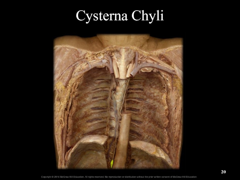
Saclike cisterna
chyli at its baseReceives lipid-rich chyle from GI tract
Drains lymph from left side of head and neck, left upper limb, left side of thorax, abdomen, and both lower limbs
21.1b Lymphatic Vessels, Trunks, and Ducts
Figure 21.3b
Слайд 21Clinical View: Lymphedema
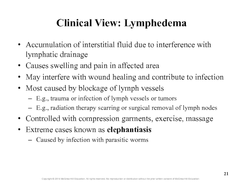
Accumulation of interstitial fluid due to interference with
lymphatic drainage
Causes swelling and pain in affected area
May interfere with
wound healing and contribute to infectionMost caused by blockage of lymph vessels
E.g., trauma or infection of lymph vessels or tumors
E.g., radiation therapy scarring or surgical removal of lymph nodes
Controlled with compression garments, exercise, massage
Extreme cases known as elephantiasis
Caused by infection with parasitic worms
Слайд 22What did you learn?
How does fluid enter lymphatic capillaries, and
what prevents its escape from them?
What is the order of
flow through different lymphatic vessels?Which lymphatic duct receives lymph from the right leg?
Слайд 23
21.2
Overview of Lymphatic Tissue and Organs
Name the two categories
of lymphatic tissue and organs, and identify components of the
body that belong to each category.Learning Objective:
Слайд 2421.2 Overview of Lymphatic Tissue and Organs
Primary lymphatic structures
Involved in
formation and maturation of lymphocytes
Red bone marrow and thymus
Secondary lymphatic
structuresDo not form lymphocytes, but house them and other immune cells
Sites of immune response initiation
Include lymph nodes, spleen, tonsils, and lymphatic nodules
Include MALT (mucosa-associated lymphatic tissue)
Слайд 25What did you learn?
What does the acronym MALT stand for?
What
is an example of a primary lymphatic structure?
Слайд 27
21.3
Overview of Lymphatic Tissue and Organs
Describe the location and
general function of red bone marrow.
Identify the two major types
of lymphocytes.Describe the structure and general function of the thymus.
Learning Objectives:
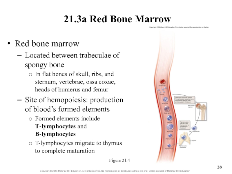
Слайд 2821.3a Red Bone Marrow
Red bone marrow
Located between trabeculae of spongy
bone
In flat bones of skull, ribs, and sternum, vertebrae, ossa
coxae, heads of humerus and femurSite of hemopoiesis: production of blood’s formed elements
Formed elements include T-lymphocytes and B-lymphocytes
T-lymphocytes migrate to thymus to complete maturation
Figure 21.4
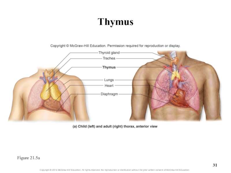
Слайд 2921.3b Thymus
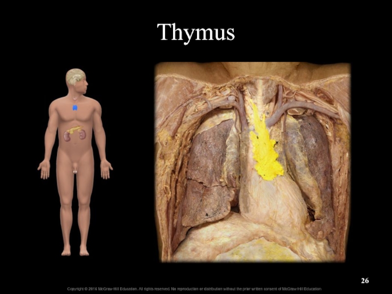
Thymus involved in T-lymphocyte maturation
Located in mediastinum
Larger in children
than adults
Grows until puberty, then regresses; gradually replaced by adipose
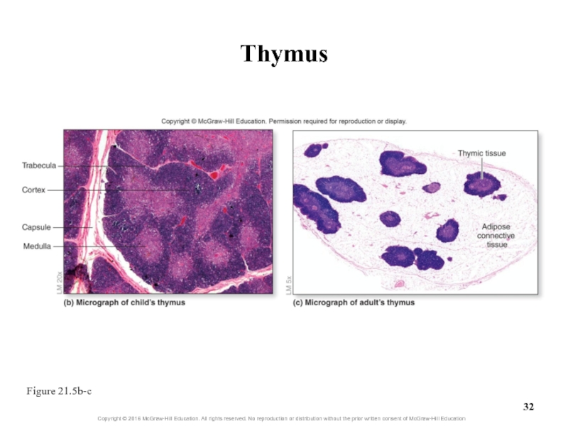
tissueConsists of two thymic lobes, each surrounded by connective tissue capsule
Trabeculae of capsules subdivide lobes into lobules
Each lobule has outer cortex and inner medulla regions
Both regions are lymphoepithelial (contain lymphatic cells and epithelial tissue)
Cortex contains immature T-lymphocytes
Medulla contains mature T-lymphocytes
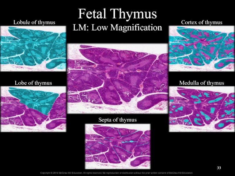
Слайд 33Fetal Thymus
LM: Low Magnification
Lobule of thymus
Lobe of thymus
Septa of thymus
Cortex
of thymus
Medulla of thymus
Слайд 35
21.4
Secondary Lymphatic Structures
Describe the structure of lymph nodes.
Explain the
function of lymph nodes.
Describe the spleen and its location.
Distinguish between
white pulp and red pulp.List the functions of the spleen.
Identify the main groups of tonsils and their location and function.
Describe the composition of individual lymphatic nodules.
Compare the locations of MALT and Peyer patches.
Learning Objectives:
Слайд 3621.4 Secondary Lymphatic Structures
Secondary lymphatic structures
House lymphocytes and other immune
cells within a reticular connective tissue matrix
Organized into lymphatic organs
and aggregates of lymphatic nodulesOrgans have a complete capsule of dense irregular connective tissue
Spleen and lymph nodes
Other lymphatic structures have an incomplete capsule or lack one
Tonsils, MALT, diffuse lymphatic nodules
Слайд 3721.4a Lymph Nodes
Lymph nodes filter lymph, remove unwanted substances
Small, oval,
encapsulated structures
Located along deep and superficial pathways of lymph vessels
Occur
in clusters receiving lymph from body regionsCervical lymph nodes receive lymph from head, neck
Axillary lymph nodes receive lymph from breast, axilla, and upper limb
Inguinal lymph nodes in groin receive lymph from lower limb and pelvis
Слайд 3821.4a Lymph Nodes
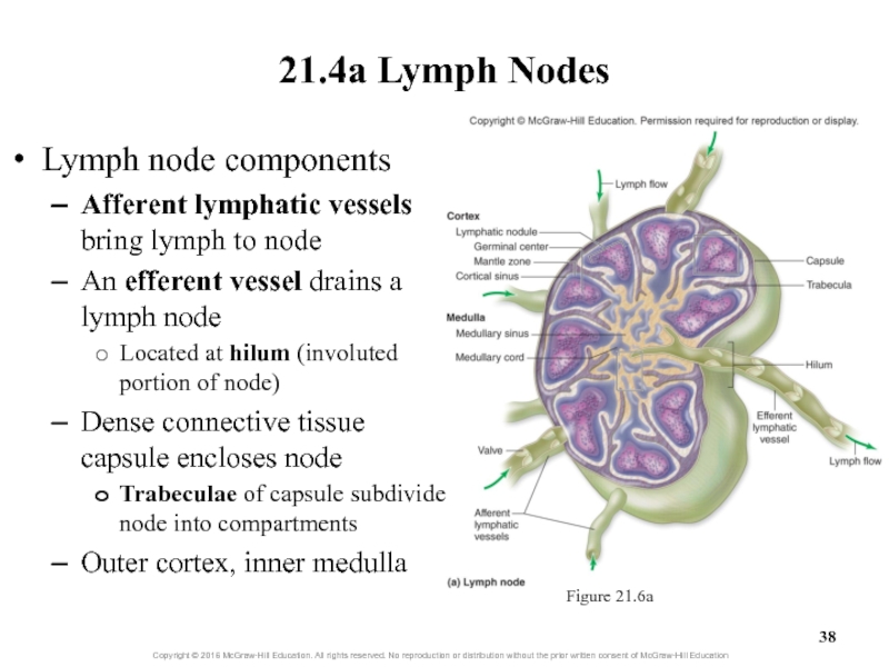
Lymph node components
Afferent lymphatic vessels bring lymph to
node
An efferent vessel drains a lymph node
Located at hilum (involuted
portion of node)Dense connective tissue capsule encloses node
Trabeculae of capsule subdivide node into compartments
Outer cortex, inner medulla
Figure 21.6a
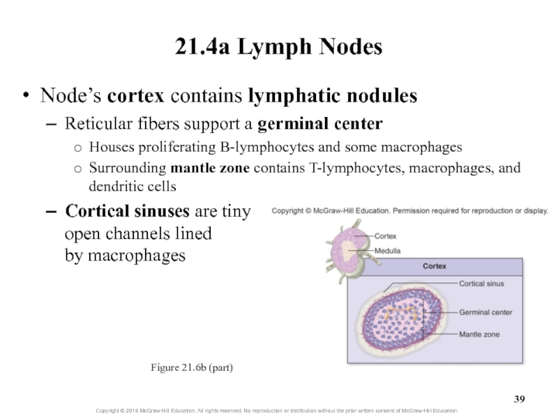
Слайд 39Node’s cortex contains lymphatic nodules
Reticular fibers support a germinal center
Houses
proliferating B-lymphocytes and some macrophages
Surrounding mantle zone contains T-lymphocytes, macrophages,
and dendritic cellsCortical sinuses are tiny open channels lined by macrophages
21.4a Lymph Nodes
Figure 21.6b (part)
Слайд 4021.4a Lymph Nodes
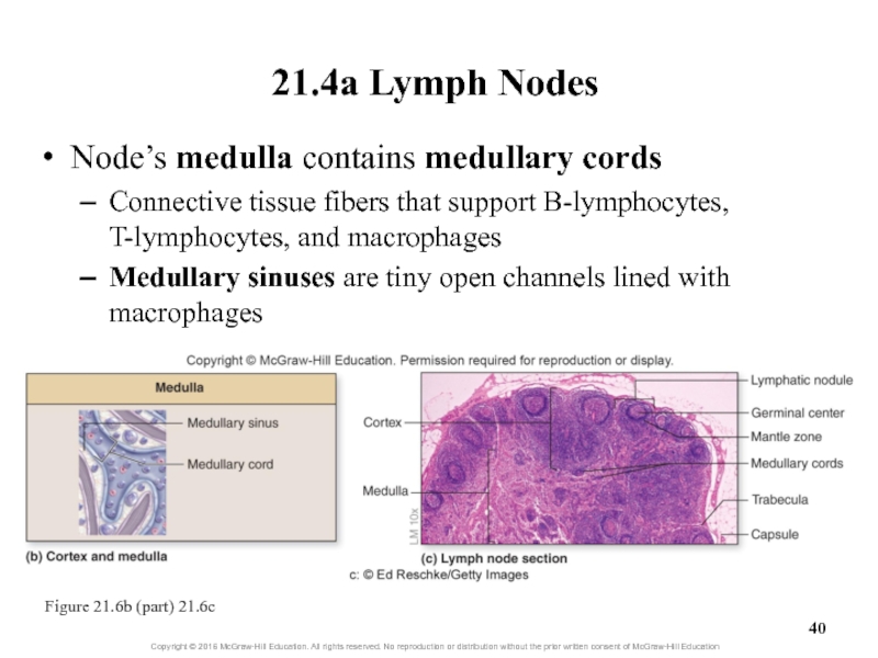
Node’s medulla contains medullary cords
Connective tissue fibers that
support B-lymphocytes, T-lymphocytes, and macrophages
Medullary
sinuses are tiny open channels lined with macrophagesFigure 21.6b (part) 21.6c
Слайд 4221.4a Lymph Nodes
Lymph flow through nodes
Lymph enters through several afferent
vessels
Creates pressure to push it through node to the single
exit vesselLymph is monitored for presence of foreign material
Macrophages remove foreign debris from lymph
Lymphocytes may initiate immune response
Lymphocytes proliferate especially in germinal centers
Can cause enlarged nodes that can be palpated in neck and axilla
Lymph exits node through efferent vessel
May enter nearby lymph node within the cluster of nodes
Слайд 43Clinical View: Lymphoma
Malignant neoplasm from lymphatic structures
Nontender, enlarged lymph node
Possible
night sweats, fever, and weight loss
Hodgkin lymphoma
Affects young adults and
people over 60If caught early, cured by excision, radiation, and chemotherapy
Non-Hodgkin lymphoma
More common
Some kinds aggressive, others slow growing
Слайд 4421.4b Spleen
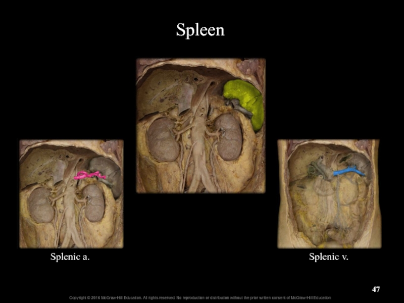
The spleen is the largest lymphatic organ
Located in left
upper abdominal quadrant
Lateral to left kidney and posterolateral to stomach
Posterolateral
aspect is convex and roundedAnteromedial border is concave
Hilum: indentation where blood vessels and nerves enter
Splenic artery supplies blood; splenic vein drains it
Surrounded by connective tissue capsule
Trabeculae from capsule extend into spleen dividing red and white pulp
Слайд 4621.4b Spleen
Tissues of the spleen
White pulp
Clusters of T- and B-lymphocytes
and macrophages around central artery
Red pulp
Contains erythrocytes, platelets, macrophages, and
B-lymphocytesSplenic cords: cells and reticular connective tissue in red pulp
Splenic sinusoids: very permeable capillaries
Storage site for erythrocytes and platelets
Слайд 50Spleen
LM: Medium Magnification
Capsule
Red pulp
White pulp
Trabeculae
Germinal center of white pulp
Central white
pulp arteries
Слайд 5121.4b Spleen
Monitoring blood as it flows through the spleen
The spleen
filters and monitors blood (not lymph)
White pulp monitors it for
foreign materials and bacteriaMacrophages in sinusoids of red pulp remove particles
Phagocytize bacteria, debris, defective erythrocytes and platelets
Path of flow: splenic artery, central artery, sinusoids, venules, splenic vein
Summary of functions: eat foreign particles, clear defective erythrocytes and platelets, store erythrocytes and platelets
In first 5 months of fetal life, spleen makes blood cells
This function can be reactivated under certain conditions
E.g., some hematologic disorders
Слайд 52Clinical View: Splenectomy
Surgical removal of the spleen
May be performed due
to
Ruptured spleen from abdominal injury (most common)
Infection, cyst, or tumor
Lymphoma
or other cancerBlood disorders (e.g., sickle cell anemia)
May be more prone to life-threatening infection
Слайд 53Tonsils
Secondary lymphatic structures
Immune surveillance of inhaled and ingested substances
Tonsillar crypts
Invaginations that trap material
Contain lymphatic nodules
Some with germinal centers
21.4c
TonsilsFigure 21.8b, c
Слайд 54Tonsils (continued)
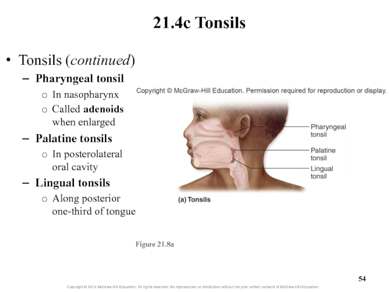
Pharyngeal tonsil
In nasopharynx
Called adenoids when enlarged
Palatine tonsils
In posterolateral
oral cavity
Lingual tonsils
Along posterior one-third of tongue
21.4c Tonsils
Figure 21.8a
Слайд 57Clinical View: Tonsillitis and Tonsillectomy
Acute tonsillitis: inflammation and infection of
tonsils
Palatine tonsils most commonly affected
Redden and enlarge
May partially obstruct pharynx
Fever,
chills, sore throat, and difficulty swallowingMay be infected by viruses or bacteria
Persistent recurrent infections, chronic tonsillitis
May require tonsillectomy, surgical removal of tonsils
Слайд 5821.4d Lymphatic Nodules and MALT
Lymphatic nodules
Clusters of lymphatic cells with
some extracellular matrix
Not completely surrounded by connective tissue capsule
Scattered nodules
termed diffuse lymphatic tissueFound in every body organ
Help defend against infection
In some areas, group together to form larger structures
E.g., MALT
Слайд 5921.4d Lymphatic Nodules and MALT
MALT
Mucosa-associated lymphatic tissue
Located in gastrointestinal, respiratory,
genital, and urinary tracts
Found in lamina propria of the mucosa
Help
defend against foreign substancesProminent in small intestines, especially ileum
Peyer patches: large collections of lymphatic nodules that form bulges in ileum wall
Слайд 62What did you learn?
What is the typical ratio of afferent
and efferent vessels at a lymph node?
Which side of the
body is the spleen on? Which direction does its hilum face?What functions does the red pulp of the spleen serve?
What are Peyer patches?

