Разделы презентаций
- Разное
- Английский язык
- Астрономия
- Алгебра
- Биология
- География
- Геометрия
- Детские презентации
- Информатика
- История
- Литература
- Математика
- Медицина
- Менеджмент
- Музыка
- МХК
- Немецкий язык
- ОБЖ
- Обществознание
- Окружающий мир
- Педагогика
- Русский язык
- Технология
- Физика
- Философия
- Химия
- Шаблоны, картинки для презентаций
- Экология
- Экономика
- Юриспруденция
Occult With No Classic CNV
Содержание
- 1. Occult With No Classic CNV
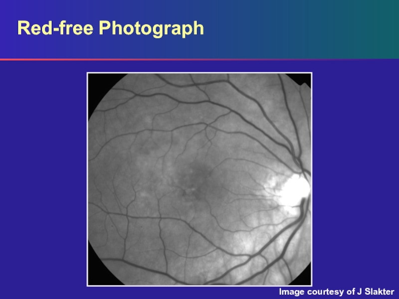
- 2. Red-free PhotographImage courtesy of J Slakter
- 3. Red-free PhotographRed-free photograph: shallow subretinal fluid and
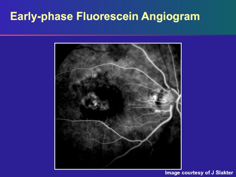
- 4. Early-phase Fluorescein AngiogramImage courtesy of J Slakter
- 5. Early-phase Fluorescein AngiogramEarly phase: an area of
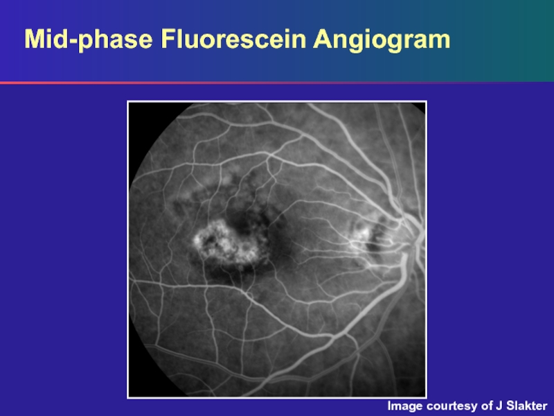
- 6. Mid-phase Fluorescein AngiogramImage courtesy of J Slakter
- 7. Mid-phase Fluorescein AngiogramMid (venous) phase: the area
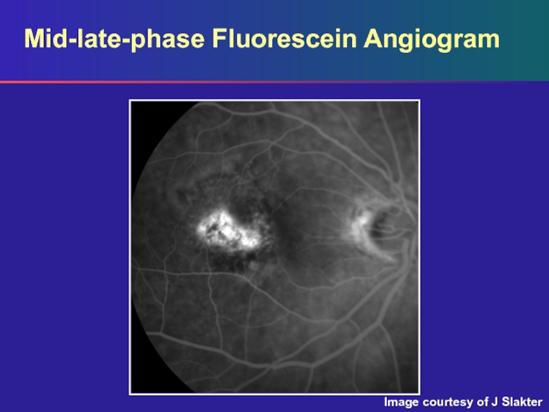
- 8. Mid-late-phase Fluorescein AngiogramImage courtesy of J Slakter
- 9. Mid-late-phase Fluorescein AngiogramMid-late phase: there is slightly more hyperfluorescence from the lesion at this point
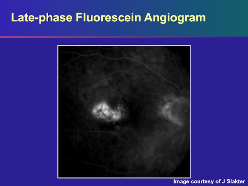
- 10. Late-phase Fluorescein AngiogramImage courtesy of J Slakter
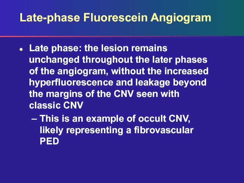
- 11. Late-phase Fluorescein AngiogramLate phase: the lesion remains
- 12. Скачать презентанцию
Red-free PhotographImage courtesy of J Slakter
Слайды и текст этой презентации
Слайд 3Red-free Photograph
Red-free photograph: shallow subretinal fluid and drusen are seen
clinical examination
Слайд 5Early-phase Fluorescein Angiogram
Early phase: an area of irregular hyperfluorescence noted
centrally with some surrounding areas of hypofluorescence
The margins of this
region are fairly well definedСлайд 7Mid-phase Fluorescein Angiogram
Mid (venous) phase: the area of hyperfluorescence stands
out in better contrast to the more homogenous background fluorescence
Note: there is no increased hyperfluorescence of the lesion typically seen with classic CNV
Слайд 9Mid-late-phase Fluorescein Angiogram
Mid-late phase: there is slightly more hyperfluorescence from
the lesion at this point
Слайд 11Late-phase Fluorescein Angiogram
Late phase: the lesion remains unchanged throughout the
later phases of the angiogram, without the increased hyperfluorescence and
leakage beyond the margins of the CNV seen with classic CNVThis is an example of occult CNV, likely representing a fibrovascular PED

