Разделы презентаций
- Разное
- Английский язык
- Астрономия
- Алгебра
- Биология
- География
- Геометрия
- Детские презентации
- Информатика
- История
- Литература
- Математика
- Медицина
- Менеджмент
- Музыка
- МХК
- Немецкий язык
- ОБЖ
- Обществознание
- Окружающий мир
- Педагогика
- Русский язык
- Технология
- Физика
- Философия
- Химия
- Шаблоны, картинки для презентаций
- Экология
- Экономика
- Юриспруденция
TIP ELEKTRONİĞİ
Содержание
- 1. TIP ELEKTRONİĞİ
- 2. 25.3 BÖLÜM
- 3. 3ELEKTROKARDİYOGRAFİK TEŞHİS VE TEDAVİ CİHAZLARI
- 4. 4Damar sertliği ve Koroner tıkanıklığın etkileriKalp kriziFelç
- 5. 5Damar sertliğini oluşturulan unsurlarYüksek kolesterol
- 6. 6Damar sertliğini oluşturulan unsurlar1) Aşırı kiloThe 1999
- 7. 7Damar sertliğini oluşturulan unsurlar2) Sigara Smoking is
- 8. 8Sigara
- 9. 9How does smoking cause heart disease? Smoking
- 10. 10What makes smoking harmful?Tobaccosmoke contains over 4000
- 11. 11Smoking increase in hematocrit
- 12. 12Smoking has dangerous effects on other parts of the bodyStroke
- 13. 13Smoking has dangerous effects on other parts of the bodyMany types of cancer (particularly lung cancer)
- 14. 14Smoking has dangerous effects on other parts of the bodyChronic bronchitis
- 15. 15Smoking has dangerous effects on other parts
- 16. 16Smoking has dangerous effects on other parts of the bodyBacak damarlarında varis oluşumu
- 17. 17Damar sertliğini oluşturulan unsurlar3) Kötü beslenmeTriglycerides içeren
- 18. 18Hangi kolesterol?Doğru beslenme
- 19. 19Damar sertliğini oluşturulan unsurlar4) Stres
- 20. 20What causes stress? exams Listening the news
- 21. 21Damar sertliğini oluşturulan unsurlar5) Hareketsiz yaşam
- 22. 22Physical activity plays an important part in
- 23. 23Damar sertliğini oluşturulan unsurlar6) Şeker hastalığı (Diabet) Açken kan şekeri (glukoz) 60-110 mg olmalıdır.
- 24. 24Damar sertliğini oluşturulan unsurlar7) Yüksek tansiyon
- 25. 25What Blood Pressure Is Considered Too High?
- 26. 26Damar sertliği ve Koroner tıkanıklıkDamar sertliği; Kalbi
- 27. 27Kan akışı
- 28. 28Plak ve kan akışı
- 29. 29Kalp atağıHeart Attack Pain Areas
- 30. 30Kalp atağı
- 31. 31Kalp atağı sonrası işlemler Anjiografi
- 32. 32Angiograph
- 33. 33Anjiografi Cardiac catheterization procedure
- 34. 34Coronary angiogram PictureThe arrow indicates a blockage in the right coronary artery
- 35. 35Electrophysiology Studies - EPS EPS, kalbin elektriksel
- 36. 36Electrophysiology Studies - EPS
- 37. 37Computed Tomography (CT) Scan CT scan is
- 38. 38Computed Tomography (CT) Scan
- 39. 39 This 3D spiral CT view is
- 40. 40Doppler ultrasound or "echo"Echocardiography
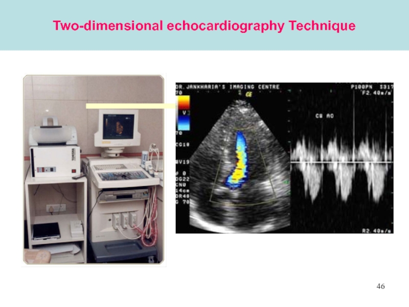
- 41. 41Echocardiography Technique
- 42. 42The echo actually consists of three different procedures:
- 43. 431) M-mode echocardiography TechniqueIn the M-mode a single beam of ultrasound is used
- 44. 44M-mode echocardiographyM-mode echocardiography provides a one-dimensional moving
- 45. 452) Two-dimensional echocardiography Technique
- 46. 46Two-dimensional echocardiography Technique
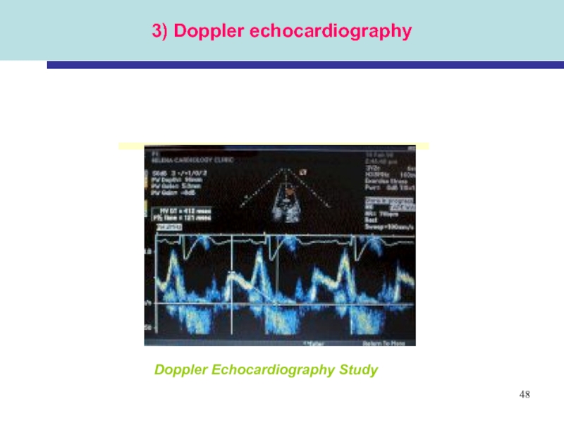
- 47. 47Echocardiography Techique3) Doppler echocardiography
- 48. 483) Doppler echocardiographyDoppler Echocardiography Study
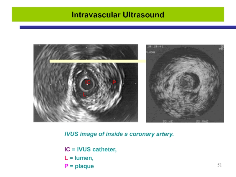
- 49. 49Intravascular Ultrasound
- 50. 50Intravascular Ultrasound
- 51. 51Intravascular Ultrasound
- 52. 52Holter Monitoring
- 53. 53Ambulatory ECG - Holter
- 54. 54Exercise EKG, or Stress Test The patient
- 55. 55Magnetic Resonance Imaging - (MRI)
- 56. 56Magnetic resonance Imaging - (MRI)MRI cihazı uzun
- 57. 57Magnetic resonance Imaging - (MRI)
- 58. 58Magnetic Resonance Imaging - (MRI)
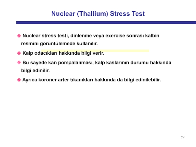
- 59. 59Nuclear (Thallium) Stress Test
- 60. 60Nuclear (Thallium) Stress Test
- 61. 61Nuclear (Thallium) Stress TestA nuclear stress test
- 62. 62Positron Emission Tomography - (PET)
- 63. 63Positron Emission Tomography - (PET)
- 64. 64Positron Emission Tomography - (PET)
- 65. 65Positron Emission Tomography - (PET)
- 66. 66Positron Emission Tomography - (PET)
- 67. 67Positron Emission Tomography - (PET)The PET scanner
- 68. 68Positron Emission Tomography - (PET)
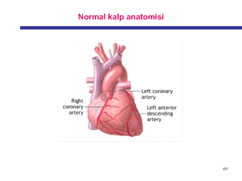
- 69. 69Normal kalp anatomisi
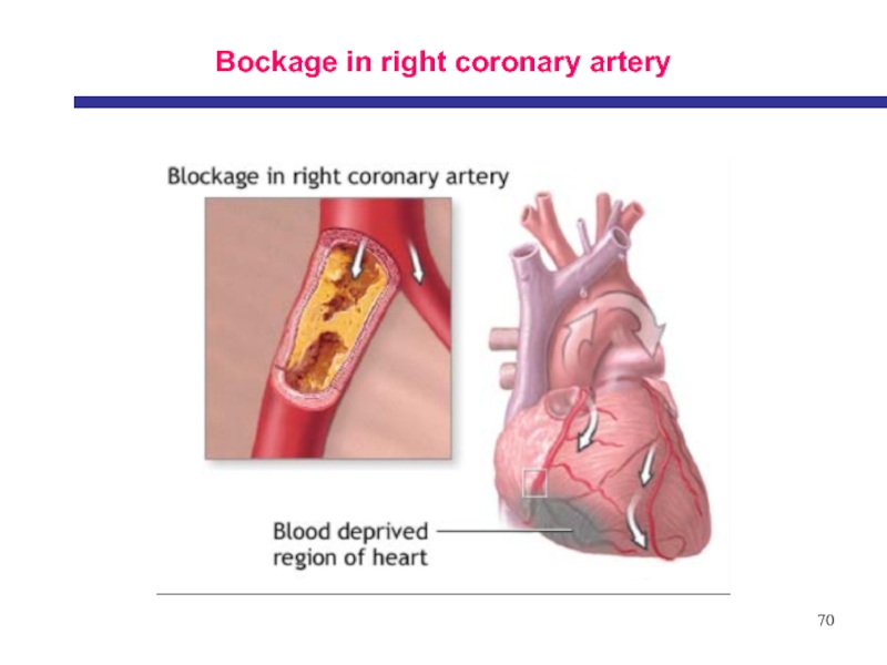
- 70. 70Bockage in right coronary artery
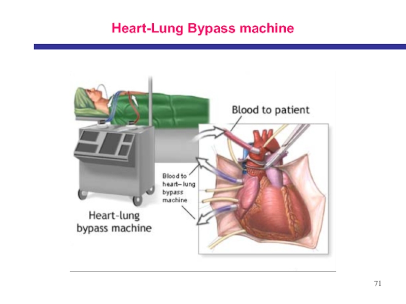
- 71. 71Heart-Lung Bypass machine
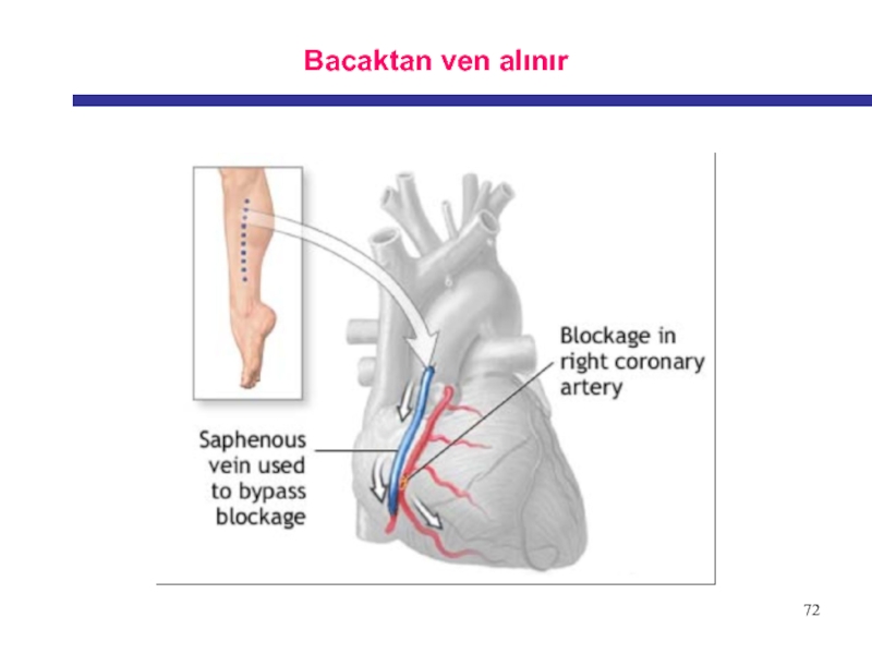
- 72. 72Bacaktan ven alınır
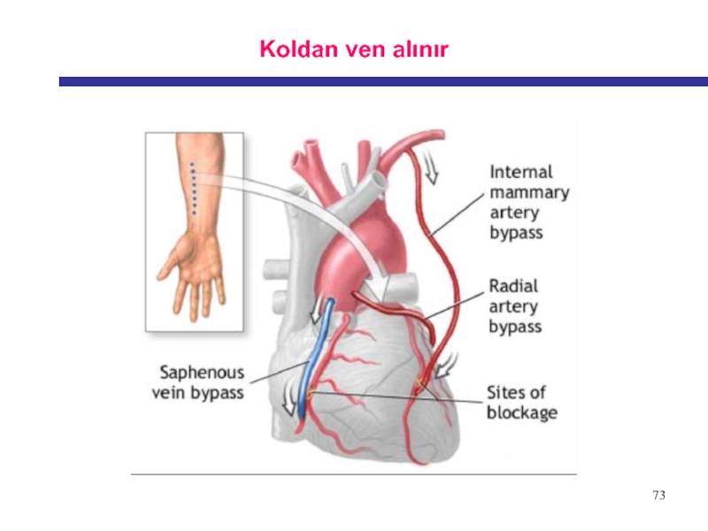
- 73. 73Koldan ven alınır
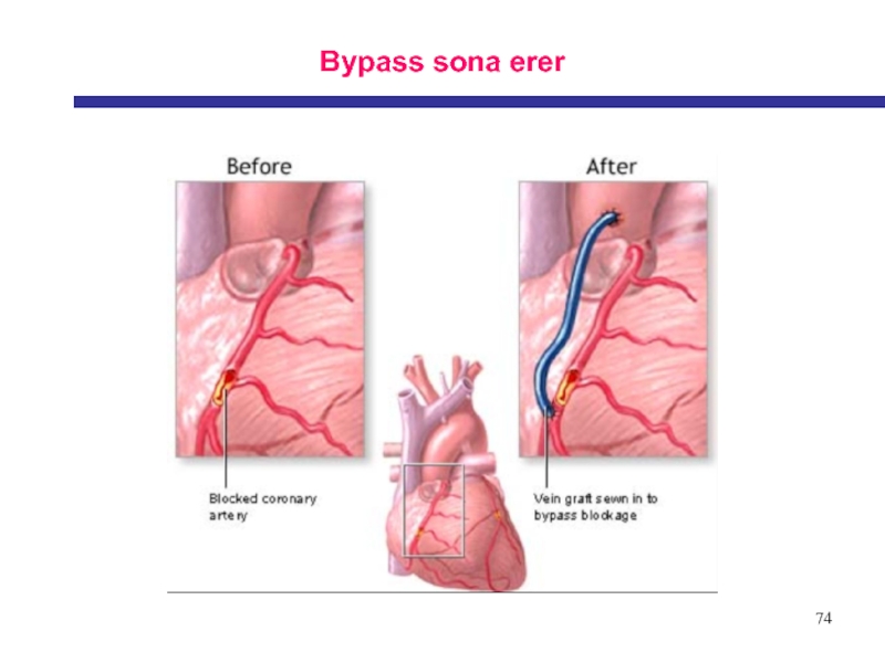
- 74. 74Bypass sona erer
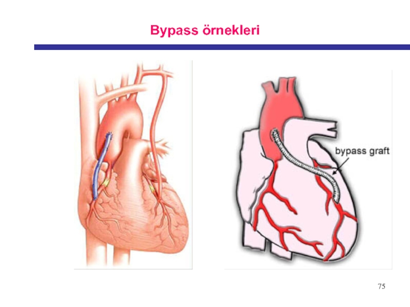
- 75. 75Bypass örnekleri
- 76. Скачать презентанцию
25.3 BÖLÜM
Слайды и текст этой презентации
Слайд 66
Damar sertliğini oluşturulan unsurlar
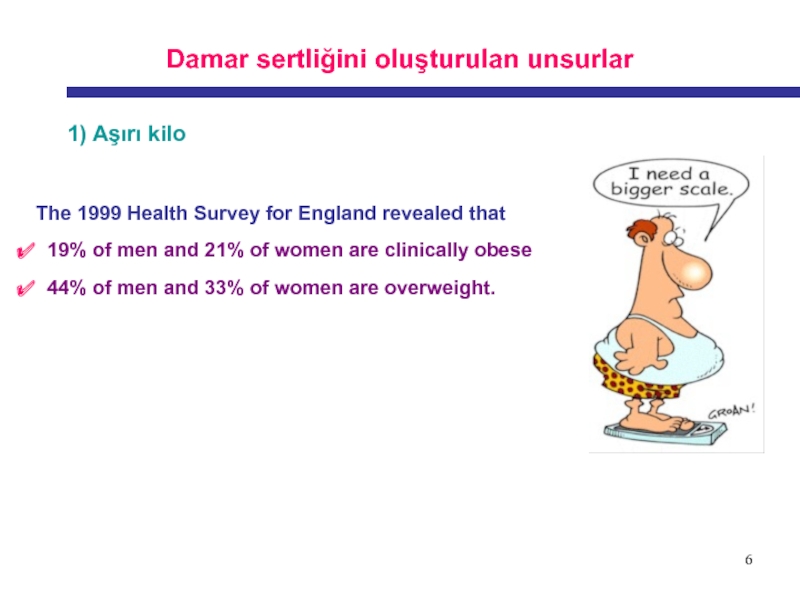
1) Aşırı kilo
The 1999 Health Survey for
England revealed that
19% of men and 21% of
women are clinically obese 44% of men and 33% of women are overweight.
Слайд 77
Damar sertliğini oluşturulan unsurlar
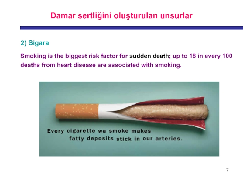
2) Sigara
Smoking is the biggest risk
factor for sudden death; up to 18 in every 100
deaths from heart disease are associated with smoking.Слайд 99
How does smoking cause heart disease?
Smoking damages coronary artery
walls eventually causing them to harden and narrow and reduce
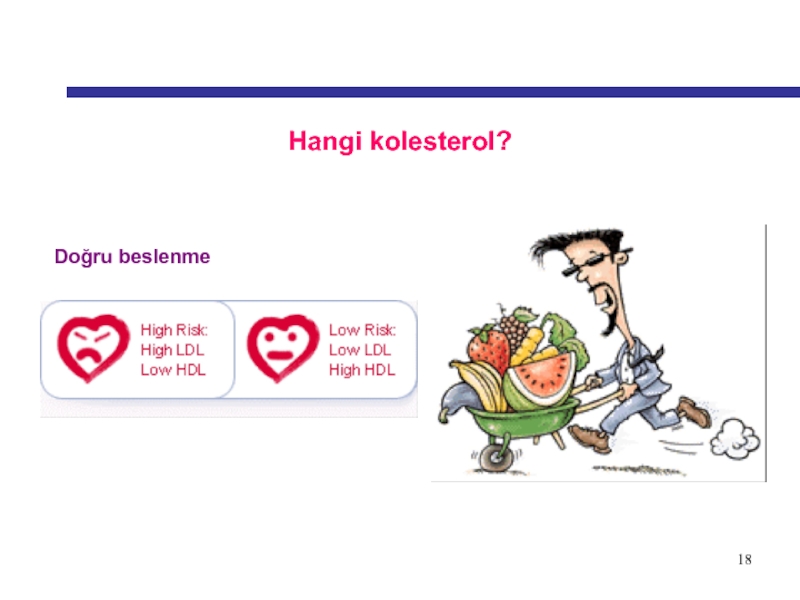
blood supply to the heart muscle.Smoking lowers HDL-cholesterol (good cholesterol).
Smoking increases fibrinogen in the blood which makes your blood more sticky and likely to clot.
Smoking increases your heart rate and blood pressure.
Smoking removes oxygen from the blood and replaces it with carbon monoxide
Слайд 1010
What makes smoking harmful?
Tobacco
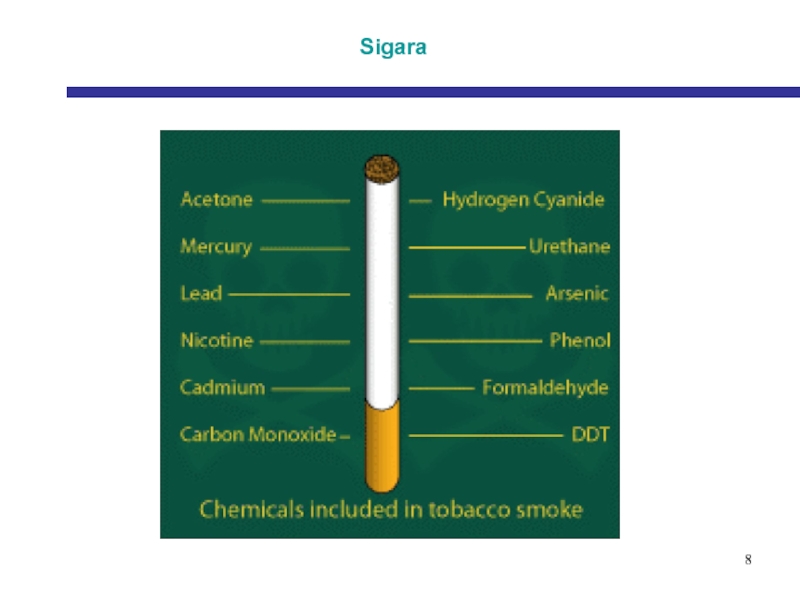
smoke contains over 4000 chemical compounds but
nicotine and carbon monoxide are the two that are most
dangerous for your heart.Nicotine
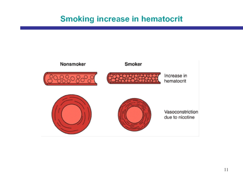
Nicotine stimulates the body to produce adrenaline which makes the heart beat faster and raises blood pressure, causing the heart to work faster.
Carbon monoxide
joins onto haemoglobin in the bloodstream making it less able to carry oxygen to the heart muscle and all other parts of the body. Up to half the blood can be carrying carbon monoxide instead of oxygen in some smokers.
Tar
is brown and treacly in appearance and causes cancer. Low tar cigarettes can be just as harmful to your heart health.
Слайд 1313
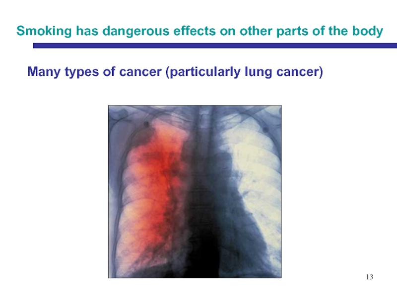
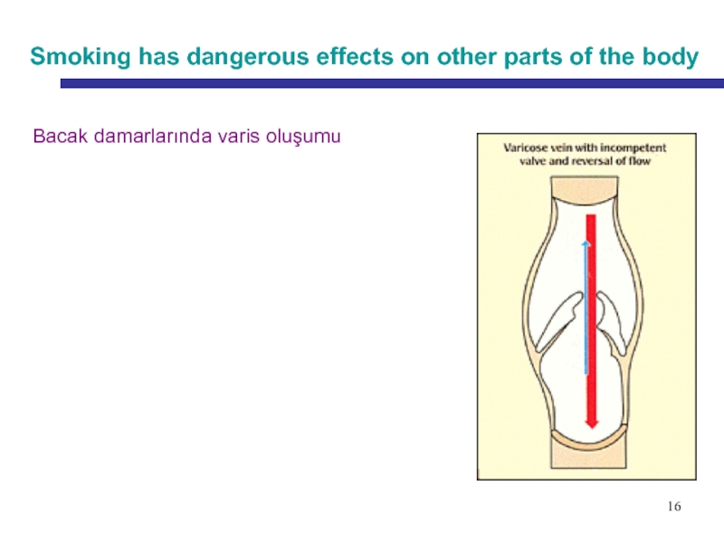
Smoking has dangerous effects on other parts of the body
Many
types of cancer (particularly lung cancer)
Слайд 1515
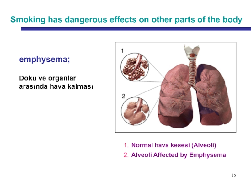
Smoking has dangerous effects on other parts of the body
emphysema;
Doku ve organlar arasında hava kalması
1. Normal hava kesesi (Alveoli)
2.
Alveoli Affected by Emphysema Слайд 1717
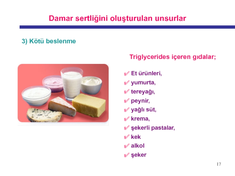
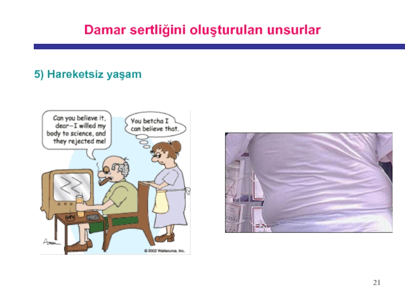
Damar sertliğini oluşturulan unsurlar
3) Kötü beslenme
Triglycerides içeren gıdalar;
Et ürünleri,
yumurta,
tereyağı,
peynir,
yağlı süt,
krema, şekerli pastalar,
kek
alkol
şeker
Слайд 2020
What causes stress?
exams Listening the news
arguments homework
being left out of a group a new school
girl friends / boy friends being late
going to the dentist moving house
taking on a new responsibility a job interview
Слайд 2222
Physical activity plays an important part in preventing heart disease
by helping to:
Reduce your risk of having a stroke.
Increase HDL-cholesterol level.
Reduce your weight
Control blood sugar (glucose).
Reduce your chance of developing diabetes.
Prevent blood clotting.
Слайд 2323
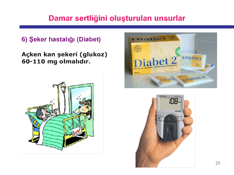
Damar sertliğini oluşturulan unsurlar
6) Şeker hastalığı (Diabet)
Açken kan şekeri
(glukoz)
60-110 mg olmalıdır.
Слайд 2525
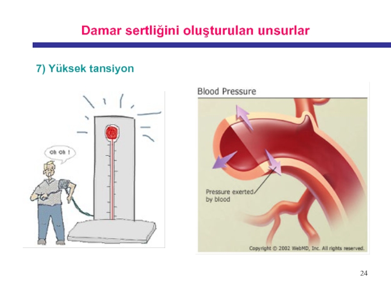
What Blood Pressure Is Considered Too High?
120/80 -
Normal
139/89 - Prehypertension
(This means that you do not have
hypertension, but you are likely to develop it in the future unless you adopt lifestyle changes to keep your blood pressure under control)140/90 - Hypertension
Слайд 2626
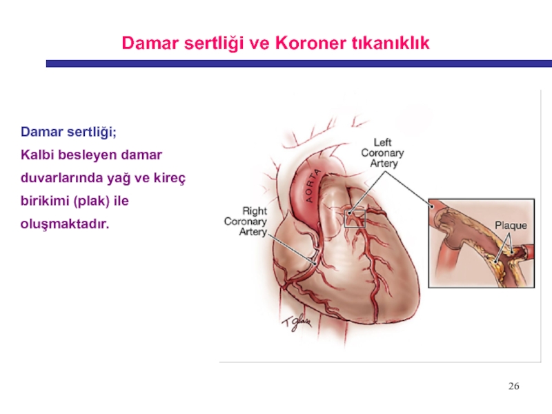
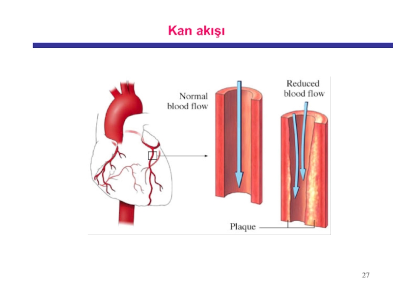
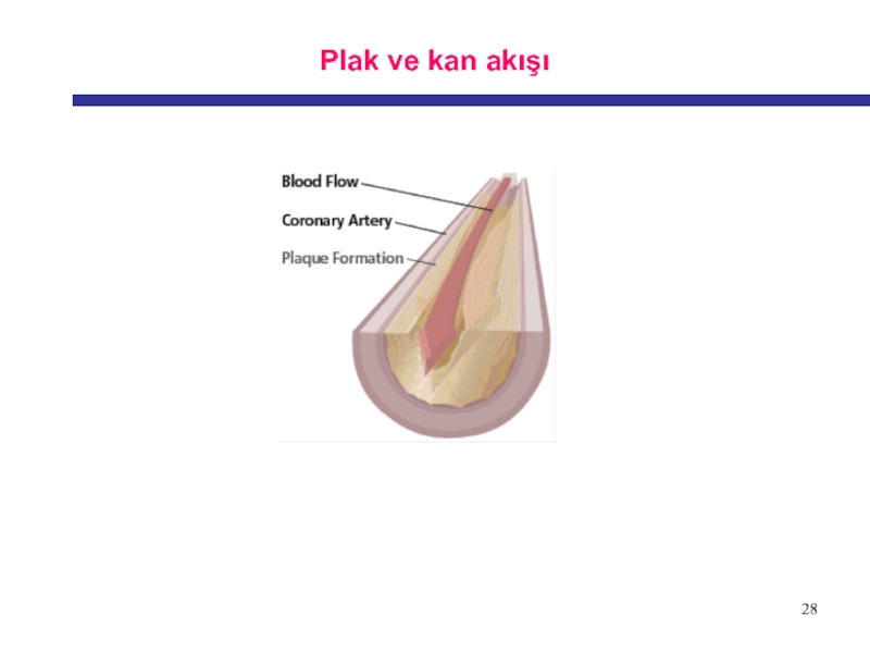
Damar sertliği ve Koroner tıkanıklık
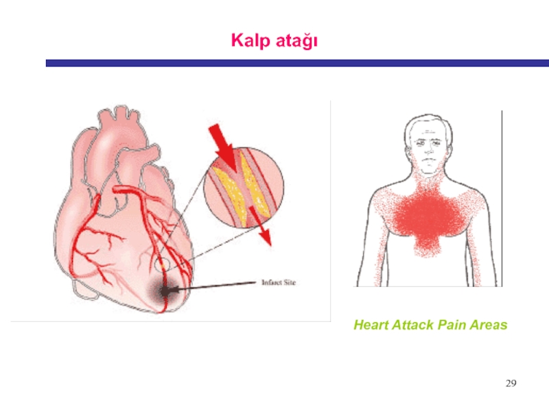
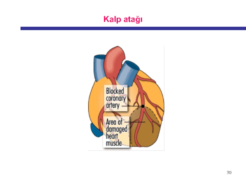
Damar sertliği;
Kalbi besleyen damar duvarlarında
yağ ve kireç birikimi (plak) ile oluşmaktadır.
Слайд 3535
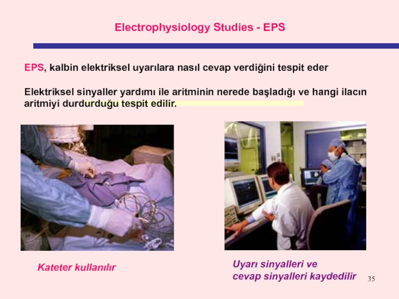
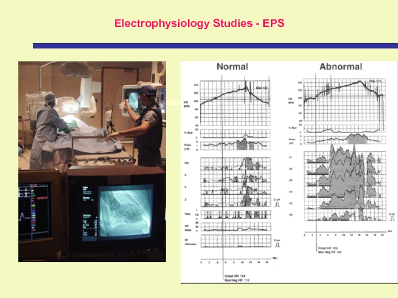
Electrophysiology Studies - EPS
EPS, kalbin elektriksel uyarılara nasıl cevap
verdiğini tespit eder Elektriksel sinyaller yardımı ile aritminin nerede başladığı ve
hangi ilacın aritmiyi durdurduğu tespit edilir.Uyarı sinyalleri ve
cevap sinyalleri kaydedilir
Kateter kullanılır
Слайд 3737
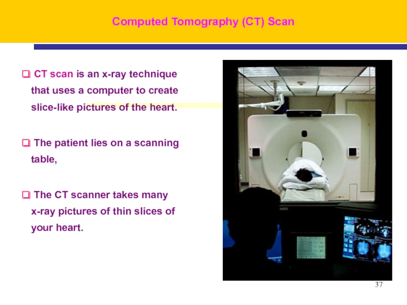
Computed Tomography (CT) Scan
CT scan is an x-ray technique
that uses a computer to create slice-like pictures of the
heart.The patient lies on a scanning table,
The CT scanner takes many x-ray pictures of thin slices of your heart.
Слайд 3939
This 3D spiral CT view is looking down on
top of the left ventricle.
It shows a normal
coronary artery (black arrow) and side branches.Computed Tomography (CT) Scan
Слайд 4444
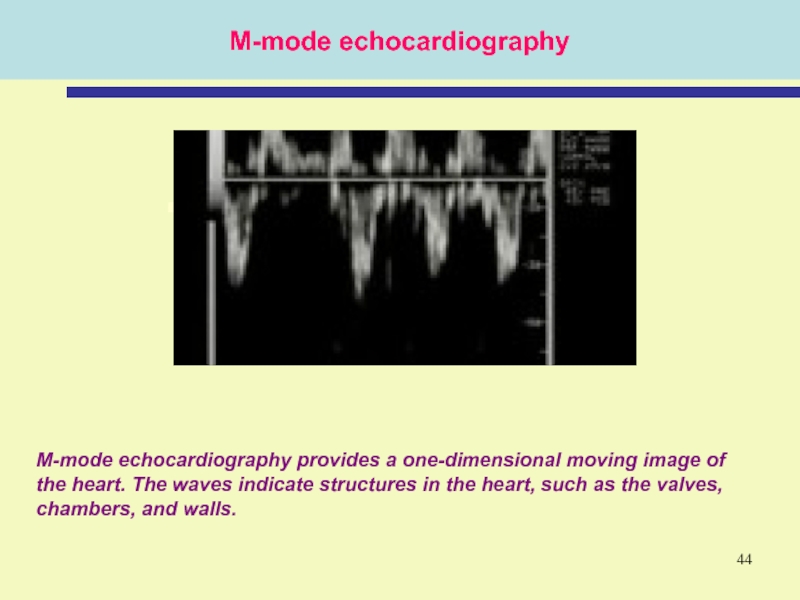
M-mode echocardiography
M-mode echocardiography provides a one-dimensional moving image of the
heart. The waves indicate structures in the heart, such as
the valves, chambers, and walls.Слайд 5454
Exercise EKG, or Stress Test
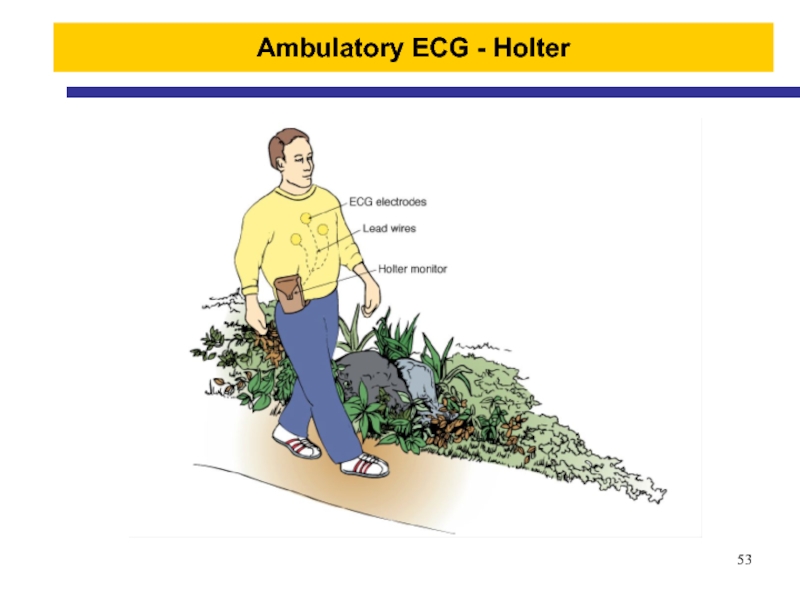
The patient is attached
to the EKG machine.
The patient exercises by walking on
a treadmill or pedaling a stationary bicycle while the EKG is recorded. This test is done to assess changes in the EKG during stress such as exercise.
Слайд 5656
Magnetic resonance Imaging - (MRI)
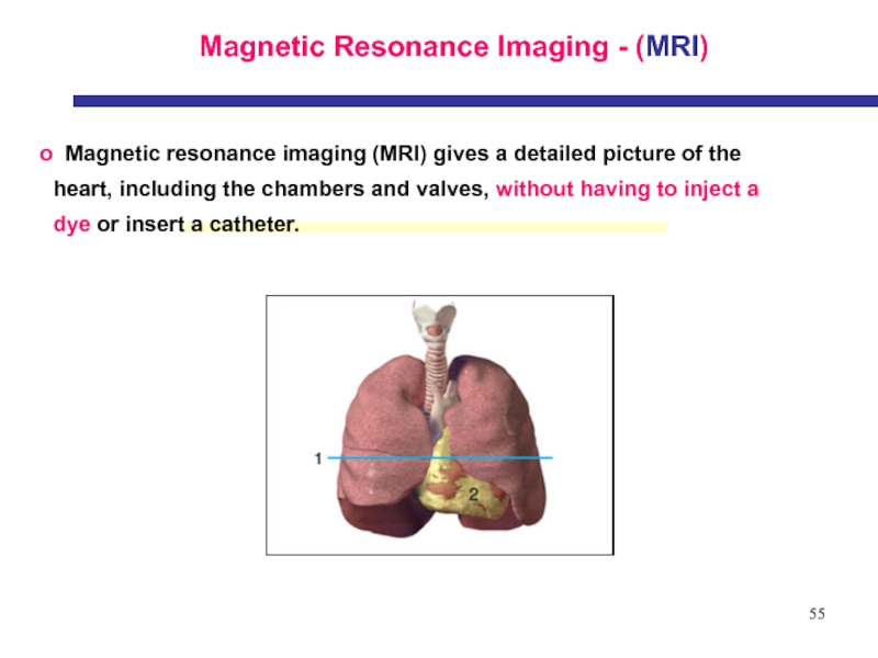
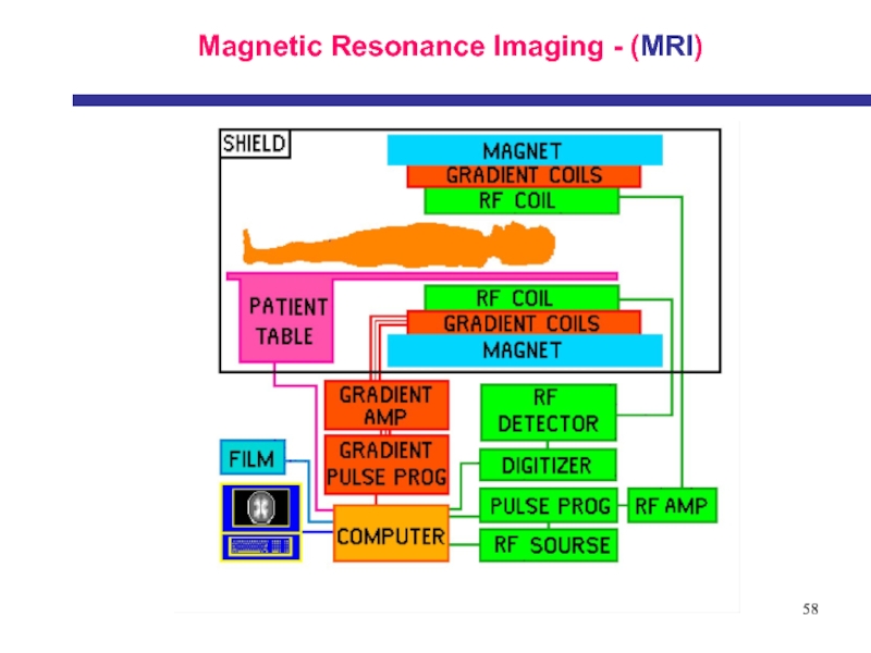
MRI cihazı uzun bir tünel veya
tüp gibidir.
Tünel içerisine hasta yatırıldığında, etrafı manyetik alanla çevrilir.
Слайд 6161
Nuclear (Thallium) Stress Test
A nuclear stress test lets
A nuclear
stress test lets
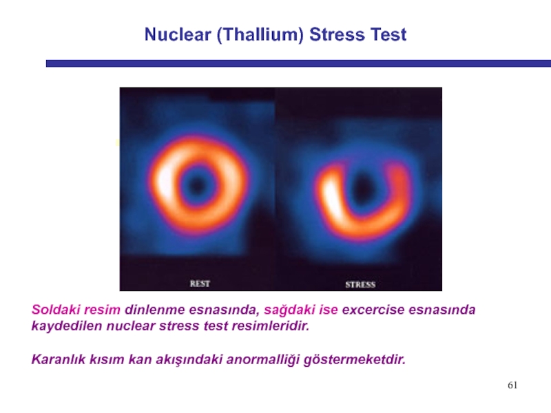
Soldaki resim dinlenme esnasında, sağdaki ise excercise
esnasında kaydedilen nuclear stress test resimleridir.Karanlık kısım kan akışındaki anormalliği göstermeketdir.
