Слайд 1Introduction to the Nervous System
Слайд 2Nervous Tissue
Body perceives and responds to multiple sensations
Controls multiple
muscle movements
Others movements without voluntary input
e.g., beating of the heart
Nervous
System
Controls and interprets all these sensations and muscle movements
Слайд 3Introduction to the Nervous System:
General Functions
Nervous system
Body’s primary communication
and control system
Integrates and regulates body functions
Uses electrical activity
transmitted
along specialized nervous system cells
Слайд 4Introduction to the Nervous System:
General Functions
Nervous system activities
Collects
information
specialized nervous structures, receptors
monitor changes in external and internal environment,
stimuli
e.g., receptors in the skin detecting information about touch
Processes and evaluates information
then determines if response required
Слайд 5Introduction to the Nervous System:
General Functions
The nervous system activities
(continued)
Initiates response to information
initiate response via nerves to effectors
include muscle
tissue and glands
e.g., muscle contraction or change in gland secretion
Слайд 6Introduction to the Nervous System:
General Functions
Structural organization: central versus
peripheral nervous system
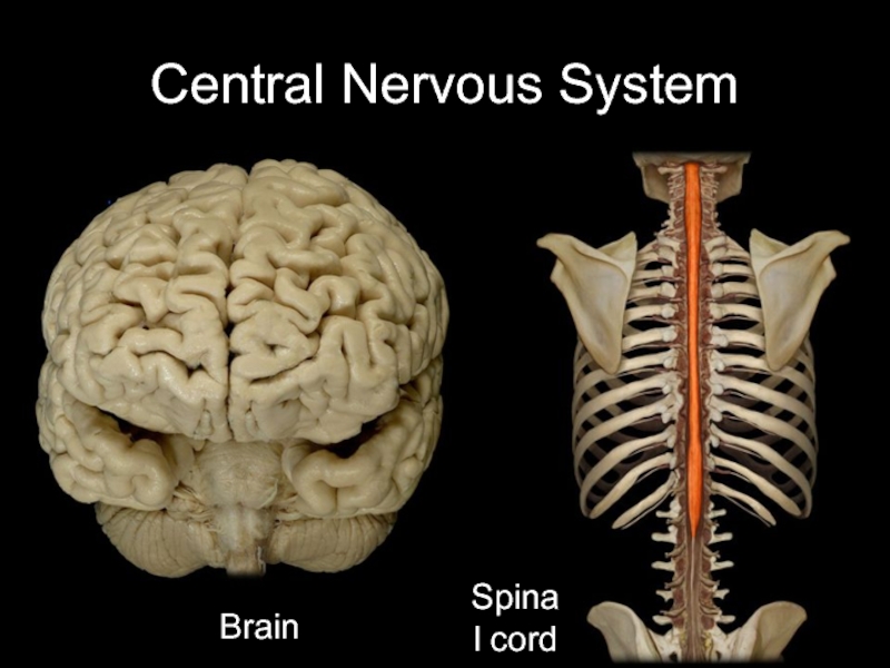
Central nervous system
anatomic division of the nervous system
includes
brain and spinal cord
brain protected in the skull
spinal cord protected in the vertebral canal
Peripheral nervous system
other anatomic division
includes nerves, bundles of neuron processes
includes ganglia, clusters of neuron cell bodies
Слайд 7Central Nervous System
Brain
Spinal cord
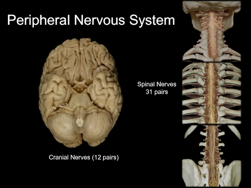
Слайд 8Peripheral Nervous System
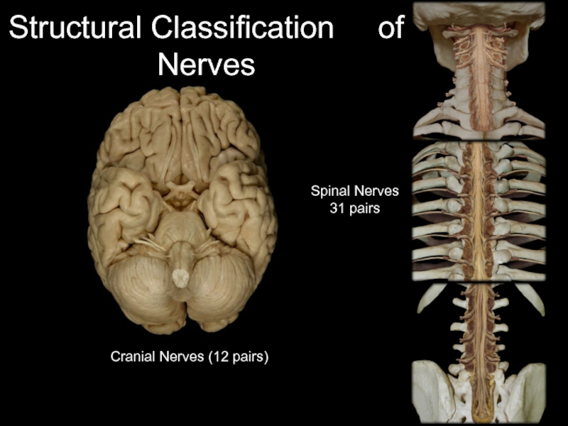
Cranial Nerves (12 pairs)
Spinal Nerves
31 pairs
Слайд 10Introduction to the Nervous System:
General Functions
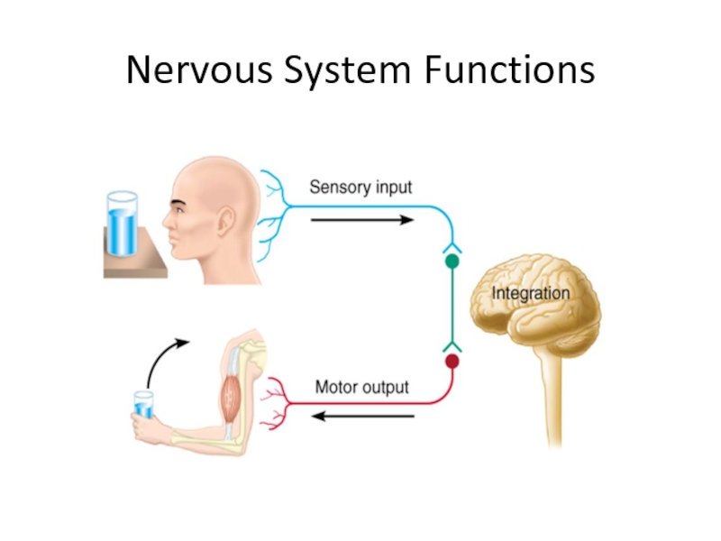
Functional organization: sensory versus
motor nervous system
Sensory nervous system
also known as afferent nervous system
responsible
for receiving sensory information from receptors
transmits information to the CNS
further divided into somatic and visceral sensory
Слайд 11Introduction to the Nervous System:
General Functions
Functional organization: sensory versus
motor nervous system (continued)
Somatic sensory
detects stimuli that we consciously perceive
receptors
include:
eyes and nose
tongue and ears
skin
proprioceptors (receptors detecting body position)
Слайд 12Introduction to the Nervous System:
General Functions
Functional organization: sensory versus
motor nervous system (continued)
Visceral sensory
detects stimuli we do not consciously
perceive
receptors include:
structures within blood vessels
structures within internal organs
e.g., detecting stretch of organ wall
Слайд 13Introduction to the Nervous System:
General Functions
Functional organization: sensory versus
motor nervous system (continued)
Motor nervous system
also known as efferent nervous
system
initiates and transmits motor output from CNS
transmits information to the effectors
may be further divided into the somatic and visceral parts
Слайд 14Introduction to the Nervous System:
General Functions
Functional organization: sensory versus
motor nervous system (continued)
Somatic motor
transmits motor output from CNS to
voluntary skeletal muscles
effector consciously controlled
e.g., pressing on accelerator of your car
Autonomic motor
transmits output from CNS without conscious control
transmits to cardiac muscle, smooth muscle, glands
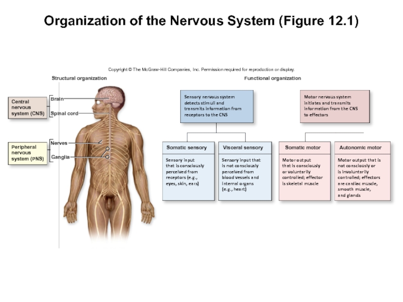
Слайд 15Organization of the Nervous System (Figure 12.1)
Copyright © The McGraw-Hill
Companies, Inc. Permission required for reproduction or display.
Motor nervous system
initiates
and transmits
information from the CNS
to effectors
Sensory input
that is consciously
perceived from
receptors (e.g.,
eyes, skin, ears)
Sensory input that
is not consciously
perceived from
blood vessels and
internal organs
(e.g., heart)
Sensory nervous system
detects stimuli and
transmits information from
receptors to the CNS
Motor output
that is consciously
or voluntarily
controlled; effector
is skeletal muscle
Motor output that is
not consciously or
is involuntarily
controlled; effectors
are cardiac muscle,
smooth muscle,
and glands
Central
nervous
system (CNS)
Peripheral
nervous
system (PNS)
Ganglia
Nerves
Brain
Spinal cord
Structural organization
Functional organization
Autonomic motor
Somatic motor
Visceral sensory
Somatic sensory
Слайд 16Introduction to the Nervous System:
General Functions
Sensory nervous system and
the motor nervous system
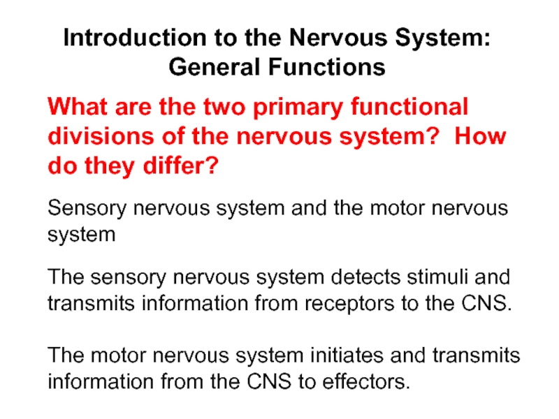
What are the two primary functional divisions
of the nervous system? How do they differ?
The sensory nervous system detects stimuli and transmits information from receptors to the CNS.
The motor nervous system initiates and transmits information from the CNS to effectors.
Слайд 17Nervous Tissue: Neurons
Two cell types in nervous tissue
Neurons
basic structural unit
of the nervous system
excitable cells that transmit electrical signals
Glial cells
nonexcitable
cells that primarily support and protect neurons
Слайд 18Neurons
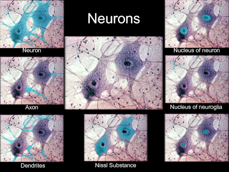
Neuron
Axon
Dendrites
Nissl Substance
Nucleus of neuron
Nucleus of neuroglia
Слайд 19Nervous Tissue—Neurons:
General Characteristics
Special characteristics of neurons
Excitability
responsive to stimulation
type dependent
on its location
most respond only to binding of molecules, neurotransmitters
Conductivity
electrical
charges propagated along membrane
can be local and short-lived or self-propagating
Слайд 20Nervous Tissue—Neurons:
General Characteristics
Special characteristics of neurons (continued)
Secretion
release neurotransmitters in
response to electrical charges
given neuron releasing only one type of
neurotransmitter
may have excitatory or inhibitory effect on target
Extreme longevity
most formed before birth still present in advanced age
Amitotic
mitotic activity lost in most neurons
not always the case (e.g., occasionally in hippocampus)
Слайд 21Nervous Tissue—Neurons:
General Characteristics
Excitability, conductivity, secretion, extreme longevity, amitotic
Name the
five distinguishing characteristics of all neurons.
Слайд 22Nervous Tissue—Neurons:
General Characteristics
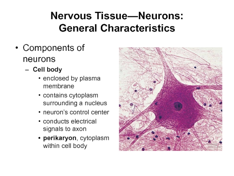
Components of neurons
Cell body
enclosed by plasma membrane
contains
cytoplasm surrounding a nucleus
neuron’s control center
conducts electrical signals to axon
perikaryon,
cytoplasm within cell body
Слайд 23Neuron Structure
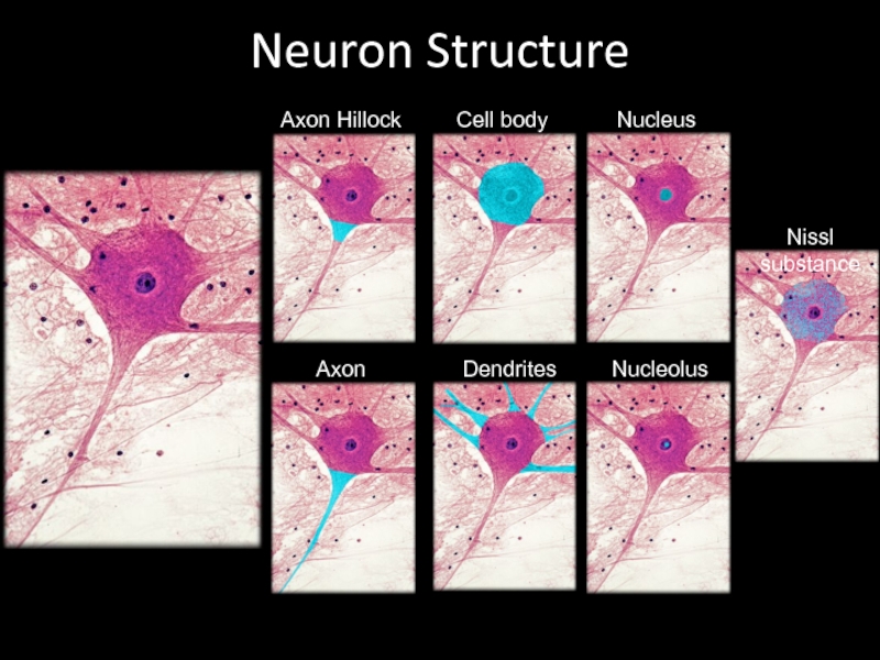
Axon Hillock
Cell body
Nucleus
Axon
Dendrites
Nucleolus
Nissl substance
Слайд 24Nervous Tissue—Neurons:
General Characteristics
Components of neurons (continued)
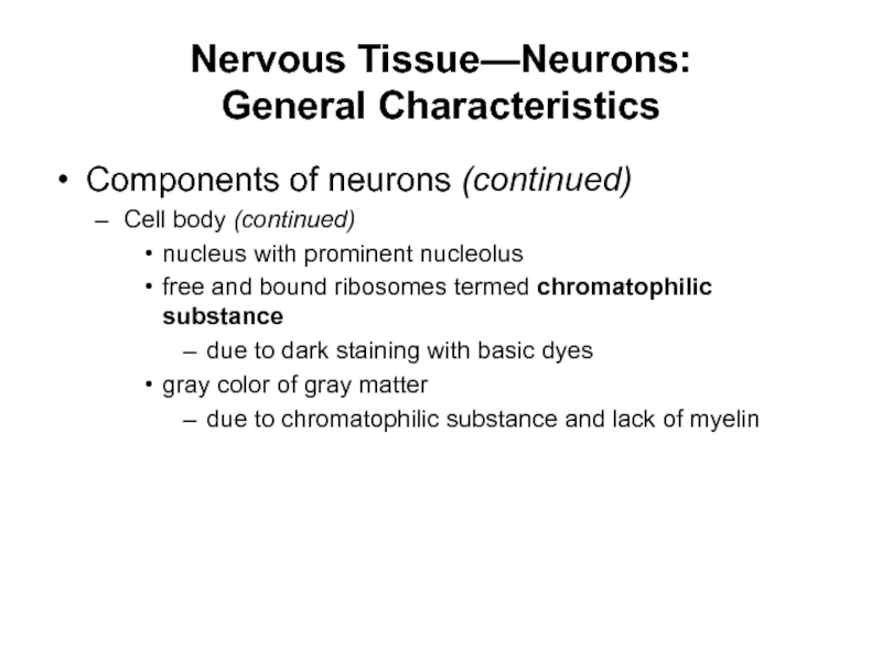
Cell body (continued)
nucleus with
prominent nucleolus
free and bound ribosomes termed chromatophilic substance
due to dark
staining with basic dyes
gray color of gray matter
due to chromatophilic substance and lack of myelin
Слайд 25Nervous Tissue—Neurons:
General Characteristics
Components of neurons (continued)
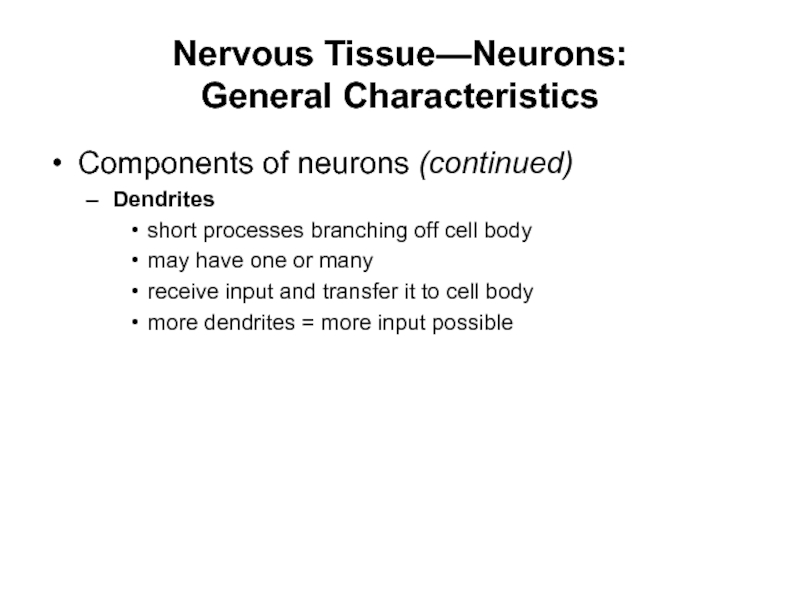
Dendrites
short processes branching off
cell body
may have one or many
receive input and transfer it
to cell body
more dendrites = more input possible
Слайд 26Nervous Tissue—Neurons:
General Characteristics
Components of neurons (continued)
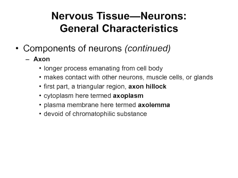
Axon
longer process emanating from
cell body
makes contact with other neurons, muscle cells, or glands
first
part, a triangular region, axon hillock
cytoplasm here termed axoplasm
plasma membrane here termed axolemma
devoid of chromatophilic substance
Слайд 27Nervous Tissue—Neurons:
General Characteristics
Components of neurons
Axon (continued)
gives rise to side
branches, axon collaterals
branch extensively at distal end into telodendria (axon
terminals)
at extreme tips, expanded regions, synaptic knobs
knobs containing numerous synaptic vesicles
contain neurotransmitter
Слайд 28Nervous Tissue—Neurons:
General Characteristics
Cytoskeleton
Composed of microfilaments, intermediate filaments, microtubules
Intermediate filaments,
termed neurofilaments
aggregate to form bundles, neurofibrils
provide tensile strength through the
neuron
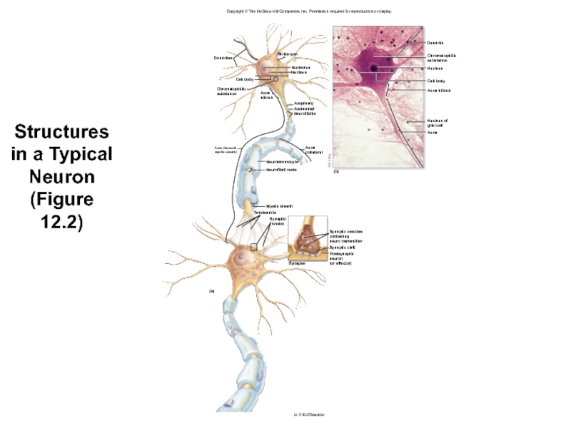
Слайд 29Structures in a Typical Neuron
(Figure
12.2)
Copyright © The McGraw-Hill Companies,
Inc. Permission required for reproduction or display.
Dendrite
Chromatophilic
substance
Nucleus
Cell body
Axon hillock
Nucleus of
glial
cell
Axon
Dendrites
Perikaryon
Axon (beneath
myelin sheath)
Chromatophilic
substance
Cell body
Axon
hillock
Neurolemmocyte
Neurofibril node
(b)
LM 100x
Axon
collateral
Nucleolus
Nucleus
Axoplasm
Axolemma
Neurofibrils
Synaptic vesicles
containing
neuro transmitter
Synaptic cleft
Postsynaptic neuron
(or effector)
Synapse
(a)
Myelin sheath
Telodendria
Synaptic
knobs
b: © Ed Reschke
Слайд 30Nervous Tissue—Neurons:
General Characteristics
Dendrites conduct electrical signals toward the cell
body. They receive input that they transfer to the cell
body.
What are the functions of dendrites, axon, and neurofibrils?
The axon is used to make contact with other neurons, muscle cells, or gland cells.
Neurofibrils give tensile support to neurons.
Слайд 31Nervous Tissue—Neurons:
Classification of Neurons
Structural classification
Structural classification of neurons
according
to number of neuron processes
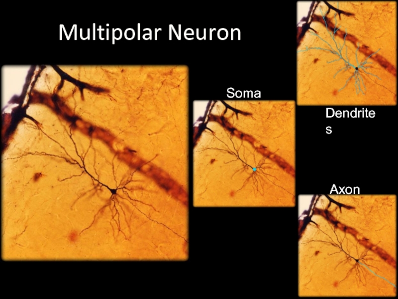
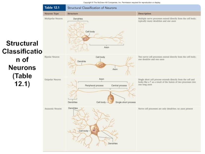
Multipolar neurons
most common type
have many dendrites
and a single axon
Bipolar neurons
have two processes extending from cell body
one dendrite and one axon
limited, e.g., in retina of the eye
Слайд 32Soma
Dendrites
Axon
Multipolar Neuron
Слайд 33Nervous Tissue—Neurons:
Classification of Neurons
Structural classification (continued)
Unipolar neurons
have single short
neuron process
emerges from cell and branches like a T
also called
pseudounipolar
start out as bipolar neurons during development
axons with peripheral process (dendrites to cell body)
axons with central process (cell body into CNS)
Слайд 34Nervous Tissue—Neurons:
Classification of Neurons
Structural classification (continued)
Anaxonic neurons
have dendrites and
no axons
produce local electrical changes but no action potentials
Слайд 35Structural Classification of Neurons
(Table
12.1)
Слайд 36Nervous Tissue—Neurons:
Classification of Neurons
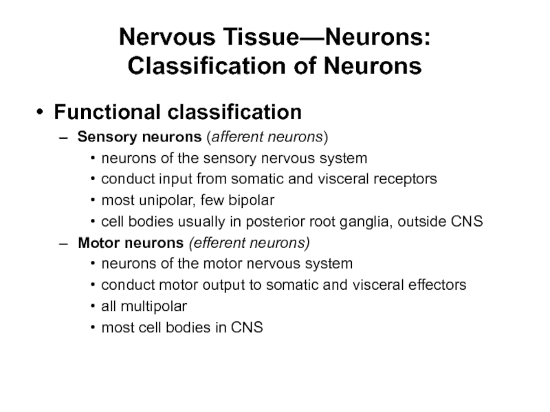
Functional classification
Sensory neurons (afferent neurons)
neurons of
the sensory nervous system
conduct input from somatic and visceral receptors
most
unipolar, few bipolar
cell bodies usually in posterior root ganglia, outside CNS
Motor neurons (efferent neurons)
neurons of the motor nervous system
conduct motor output to somatic and visceral effectors
all multipolar
most cell bodies in CNS
Слайд 37Nervous Tissue—Neurons:
Classification of Neurons
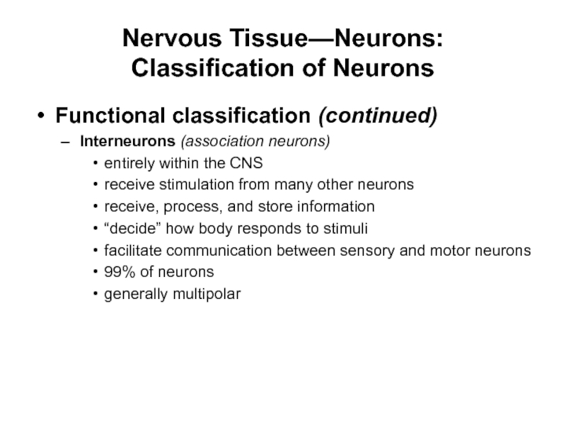
Functional classification (continued)
Interneurons (association neurons)
entirely within
the CNS
receive stimulation from many other neurons
receive, process, and store
information
“decide” how body responds to stimuli
facilitate communication between sensory and motor neurons
99% of neurons
generally multipolar
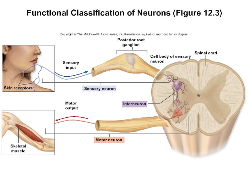
Слайд 38Functional Classification of Neurons (Figure 12.3)
Copyright © The McGraw-Hill Companies,
Inc. Permission required for reproduction or display.
Posterior root
ganglion
Spinal cord
Interneuron
Motor neuron
Skeletal
muscle
Skin
receptors
Sensory
input
Cell body of sensory
neuron
Sensory neuron
Motor
output
Слайд 39Nervous Tissue—Neurons:
Classification of Neurons
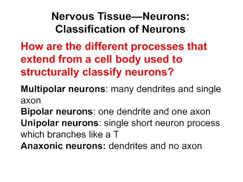
Multipolar neurons: many dendrites and single
axon
Bipolar neurons: one dendrite and one axon
Unipolar neurons: single short
neuron process which branches like a T
Anaxonic neurons: dendrites and no axon
How are the different processes that extend from a cell body used to structurally classify neurons?
Слайд 40Nervous Tissue—Neurons:
Relationship of Neurons and Nerves
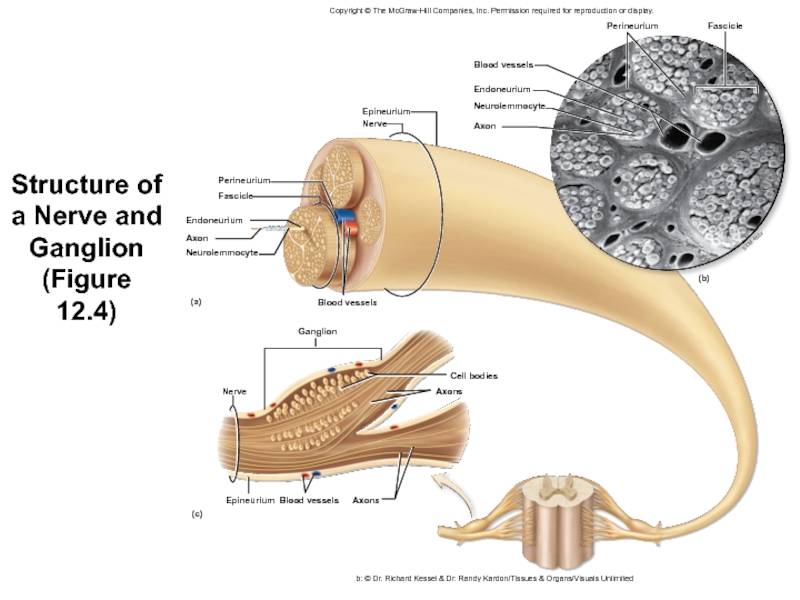
Nerve
Cablelike bundle of parallel
axons
Macroscopic structure
Epineurium
thick layer of dense irregular connective tissue
encloses the entire
nerve
provides support and protection
Слайд 41Nervous Tissue—Neurons:
Relationship of Neurons and Nerves
Nerve (continued)
Perineurium
layer of dense
irregular connective tissue
wraps bundles of axons, fascicles
supports blood vessels
Endoneurium
delicate layer
of areolar connective tissue
separates and electrically insulates each axon
has capillaries that supply the axon
Слайд 42Structure of a Nerve and Ganglion
(Figure
12.4)
Copyright © The McGraw-Hill
Companies, Inc. Permission required for reproduction or display.
b: © Dr.
Richard Kessel & Dr. Randy Kardon/Tissues & Organs/Visuals Unlimited
Fascicle
Perineurium
Epineurium
Nerve
Perineurium
Fascicle
Endoneurium
Axon
Neurolemmocyte
(a)
(c)
(b)
Ganglion
Blood vessels
Blood vessels
Endoneurium
Neurolemmocyte
Axon
Cell bodies
Axons
Nerve
Epineurium
Blood vessels
Axons
SEM 450x
Слайд 43Nervous Tissue—Neurons:
Relationship of Neurons and Nerves
Classification of nerves
Structural classification
Cranial
nerves
extend from brain
Spinal nerves
extend from spinal cord
Слайд 44Structural Classification of Nerves
Cranial Nerves (12 pairs)
Spinal Nerves
31
pairs
Слайд 45Nervous Tissue—Neurons:
Relationship of Neurons and Nerves
Classification of nerves (continued)
Functional
classification
Sensory nerves
contain only sensory neurons
Motor nerves
contain primarily motor neurons
Mixed nerves
contain
both sensory and motor neurons
most named nerves in this category
individual neurons transmitting one type of information
Слайд 46Nervous Tissue—Neurons:
Relationship of Neurons and Nerves
The epineurium encloses the
entire nerve.
What are the three connective tissue wrappings in a
nerve, and what specific structure does each ensheathe?
The perineurium encloses bundles of axons.
The endoneurium encloses individual axons.
Слайд 47Synapses
Synapse
Where neuron functionally connected to neuron or effector
Two types: chemical
and electrical
Слайд 48
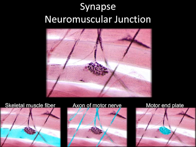
Synapse
Neuromuscular Junction
Skeletal muscle fiber
Axon of motor nerve
Motor end plate
Слайд 49Synapses
Chemical synapse
Most common
Composed of presynaptic neuron, signal producer
Composed of postsynaptic
neuron, signal receiver
Between axon and any portion of postsynaptic neuron
most
commonly with a dendrite
Knob almost touches the postsynaptic neuron
narrow fluid filled gap, the synaptic cleft
Слайд 50Neuromuscular Junction
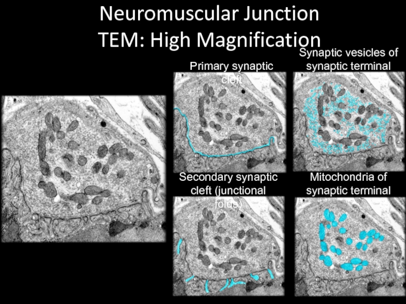
TEM: High Magnification
Primary synaptic cleft
Synaptic vesicles of synaptic
terminal
Secondary synaptic cleft (junctional folds)
Mitochondria of synaptic terminal
Слайд 51Neuromuscular Junction
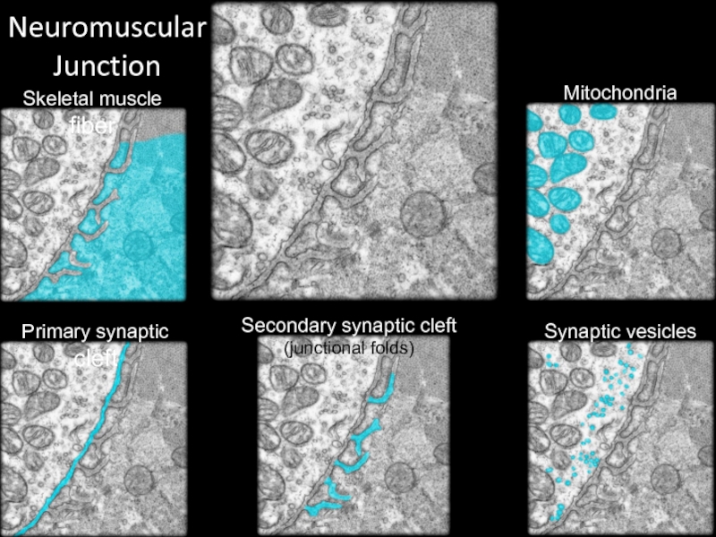
Mitochondria
Skeletal muscle fiber
Primary synaptic cleft
Synaptic vesicles
Secondary synaptic cleft (junctional
folds)
Слайд 52Synapses
Transmission at chemical synapse
Neurotransmitter molecules released from synaptic knob
Released from
synaptic vesicles into cleft
Diffusion of neurotransmitter across cleft
Binding of some
neurotransmitters to receptors
Synaptic delay
time between neurotransmitter release and binding
Single postsynaptic neuron
often stimulated by more than one neuron
Слайд 53Synapses
Electrical synapse
Much less common
Presynaptic and postsynaptic neuron physically bound together
Gap
junctions present
No delay in passing electrical signal
In limited regions of
brain and eyes
Слайд 54Synapses
Molecules stored in synaptic vesicles are released from the synaptic
knob of a presynaptic neuron into the synaptic cleft. Some
neurotransmitter diffuses across the cleft and binds receptors on the postsynaptic membrane.
What is the mode of transmission in a chemical synapse?
Слайд 55Nervous Tissue—Glial Cells:
General Characteristics
Glial cells (neuroglia)
Nonexcitable cells found in
CNS and PNS
Smaller than neurons
Capable of mitosis
Far outnumber neurons
Half volume
of nervous system
Слайд 56Nervous Tissue—Glial Cells:
General Characteristics
Glial cells (continued)
Physically protect and nourish
neurons
Provide physical scaffolding for nervous tissue
help guide migrating neurons to
their destination
Critical for normal function at neural synapses
Слайд 57Nervous Tissue—Glial Cells:
Types of Glial Cells
Glial cells of the
CNS
Astrocytes
Starlike shape from surface projections
Processes touching capillary walls and neurons
ends
termed perivascular feet
Most abundant glial cell in CNS
Слайд 58Nervous Tissue—Glial Cells:
Types of Glial Cells
Glial cells of the
CNS
Astrocytes (continued)
Help form the blood-brain barrier
feet wrap around capillaries in
the brain
together form the blood-brain barrier
strictly controls substances entering brain nervous tissue from blood
protects neurons from toxins
allows nutrients to pass
Слайд 59Nervous Tissue—Glial Cells:
Types of Glial Cells
Glial cells of the
CNS
Astrocytes (continued)
Regulate tissue fluid composition
control movement of substances between blood
and interstitial fluid
e.g., regulate K+ concentration
need constant K+ level for neuron electrical activity
Form a structural network
cytoskeleton strengthening and organizing nervous tissue
Слайд 60Nervous Tissue—Glial Cells:
Types of Glial Cells
Glial cells of the
CNS
Astrocytes (continued)
Assist neuronal development
direct development of neurons in fetal brain
secrete
chemicals regulating formation of connections
Occupy the space of dying neurons
space formerly occupied by dead neurons
filled by cells produced by astrocyte division
Слайд 61Nervous Tissue—Glial Cells:
Types of Glial Cells
Glial cells of the
CNS (continued)
Ependymal cells
Line internal cavities of brain and spinal cord
Ciliated
simple cuboidal or simple columnar epithelial cells
Slender processes with extensive branching
Form choroid plexus with nearby blood capillaries
helps produce cerebrospinal fluid
liquid that bathes external CNS and fills internal cavities
cilia helping to circulate CSF
Слайд 62Nervous Tissue—Glial Cells:
Types of Glial Cells
Glial cells of the
CNS (continued)
Microglia
Small cells with slender branches
Smallest percentage of CNS
glial cells
Phagocytic cells of the immune system
Wander CNS and replicate in infection
Engulf infectious agents
Remove debris from dead or damaged tissue
Слайд 63Nervous Tissue—Glial Cells:
Types of Glial Cells
Glial cells of the
CNS (continued)
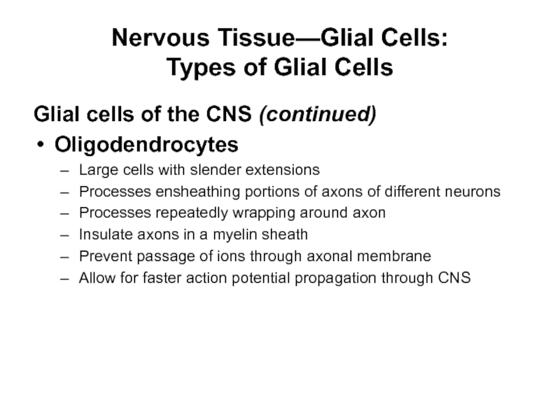
Oligodendrocytes
Large cells with slender extensions
Processes ensheathing portions of axons
of different neurons
Processes repeatedly wrapping around axon
Insulate axons in a myelin sheath
Prevent passage of ions through axonal membrane
Allow for faster action potential propagation through CNS
Слайд 64Cellular Organization of Nervous Tissue:
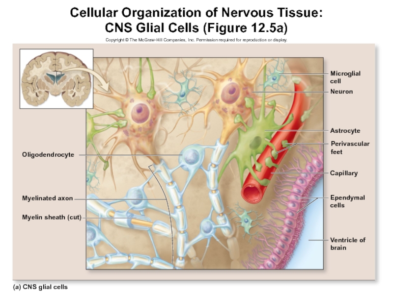
CNS Glial Cells (Figure 12.5a)
Copyright
© The McGraw-Hill Companies, Inc. Permission required for reproduction or
display.
Microglial
cell
Neuron
Astrocyte
Perivascular
feet
Capillary
Ependymal
cells
Ventricle of
brain
(a) CNS glial cells
Myelin sheath (cut)
Myelinated axon
Oligodendrocyte
Слайд 65Nervous Tissue—Glial Cells:
Types of Glial Cells
Glial cells of the
PNS
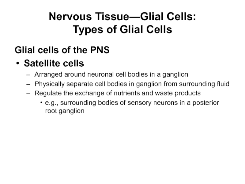
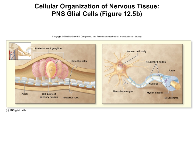
Satellite cells
Arranged around neuronal cell bodies in a ganglion
Physically separate
cell bodies in ganglion from surrounding fluid
Regulate the exchange of nutrients and waste products
e.g., surrounding bodies of sensory neurons in a posterior root ganglion
Слайд 66Nervous Tissue—Glial Cells:
Types of Glial Cells
Glial cells of the
PNS (continued)
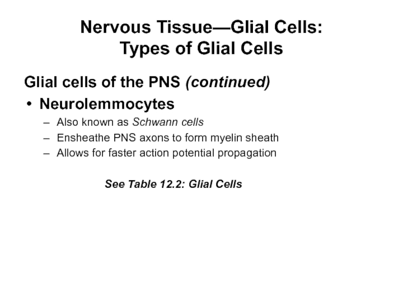
Neurolemmocytes
Also known as Schwann cells
Ensheathe PNS axons to form
myelin sheath
Allows for faster action potential propagation
See Table 12.2: Glial Cells
Слайд 67Cellular Organization of Nervous Tissue:
PNS Glial Cells (Figure 12.5b)
Copyright
© The McGraw-Hill Companies, Inc. Permission required for reproduction or
display.
Neuron cell body
Neurofibril nodes
Axon
Nucleus
Neurilemma
Myelin sheath
Neurolemmocyte
(b) PNS glial cells
Axon
Cell body of
sensory neuron
Posterior root
Posterior root ganglion
Satellite cells
Слайд 68Nervous Tissue—Glial Cells:
Types of Glial Cells
Microglia
If a person suffers
from meningitis (an inflammation of the coverings around the brain),
which type of glial cell usually replicates in response to the infection?
Слайд 69Nervous Tissue—Glial Cells:
Types of Glial Cells
Neurolemmocytes (Schwann cells)
Which specific
type of glial cell ensheathes axons in the PNS?
