Разделы презентаций
- Разное
- Английский язык
- Астрономия
- Алгебра
- Биология
- География
- Геометрия
- Детские презентации
- Информатика
- История
- Литература
- Математика
- Медицина
- Менеджмент
- Музыка
- МХК
- Немецкий язык
- ОБЖ
- Обществознание
- Окружающий мир
- Педагогика
- Русский язык
- Технология
- Физика
- Философия
- Химия
- Шаблоны, картинки для презентаций
- Экология
- Экономика
- Юриспруденция
Ministry of Education and Science of RK M. Kh. Dulaty Taraz State University
Содержание
- 1. Ministry of Education and Science of RK M. Kh. Dulaty Taraz State University
- 2. Слайд 2
- 3. Слайд 3
- 4. The human nervous system is made up
- 5. Слайд 5
- 6. BrainThe human brain serves many important functions
- 7. Brain Cells: The brain is made up
- 8. Meninges: The bony covering around the brain
- 9. The falx separates the right and left
- 10. Brainstem: The brainstem is the lower extension
- 11. Слайд 11
- 12. Thalamus: The thalamus is a structure that
- 13. Слайд 13
- 14. Cerebrum: The cerebrum forms the major portion
- 15. Слайд 15
- 16. Lobes: Lobes: Several large grooves (fissures) separate
- 17. Слайд 17
- 18. The parietal lobe processes signals received from
- 19. Hypothalamus: The hypothalamus is a structure that
- 20. Pituitary Gland: The pituitary gland is a
- 21. Скачать презентанцию
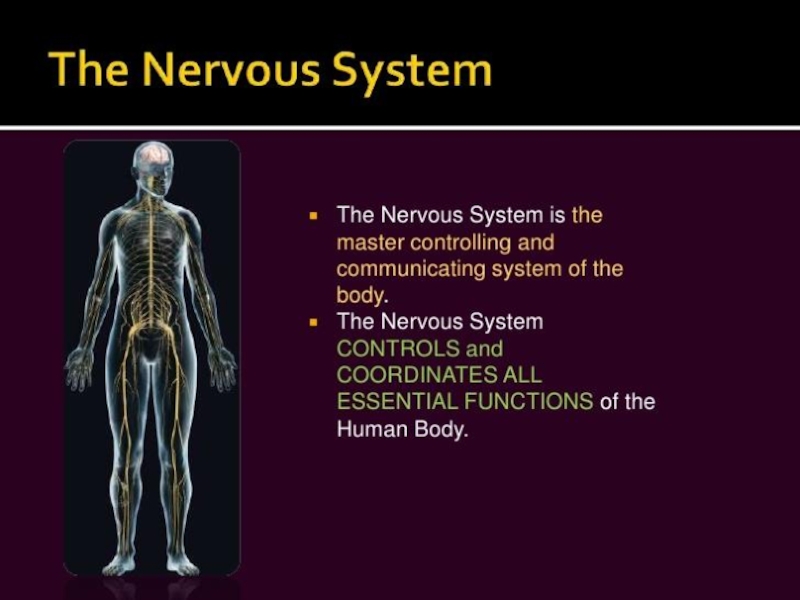
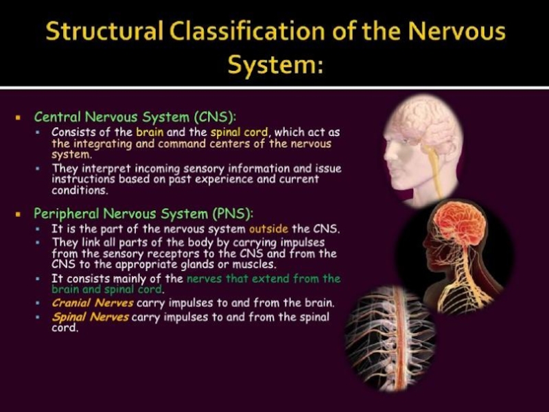
The human nervous system is made up of two main components: the central nervous system (CNS) and the peripheral nervous system (PNS). The CNS is composed of the brain, the cranial
Слайды и текст этой презентации
Слайд 1Ministry of Education and Science of RK M. Kh. Dulaty
Taraz State University Department of biology
central nervous systemСлайд 4The human nervous system is made up of two main
components: the central nervous system (CNS) and the peripheral nervous
system (PNS). The CNS is composed of the brain, the cranial nerves, and the spinal cord. The PNS is made up of the nerves that exit from the spinal cord at various levels of the spinal column as well as their tributaries.Слайд 6Brain
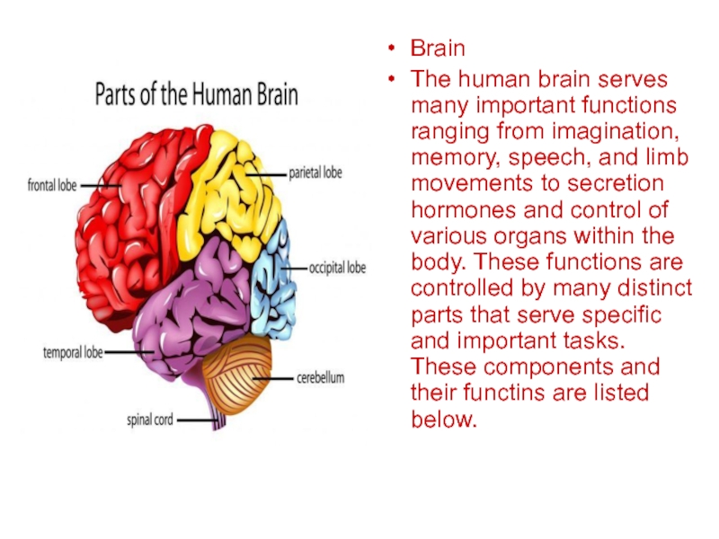
The human brain serves many important functions ranging from imagination,
memory, speech, and limb movements to secretion hormones and control
of various organs within the body. These functions are controlled by many distinct parts that serve specific and important tasks. These components and their functins are listed below.Слайд 7Brain Cells: The brain is made up of two types
of cells: neurons (yellow cells in the image below) and
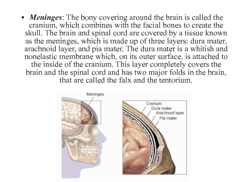
glial cells (pink and purple cells in the image below). Neurons are responsible for all of the functions that are attributed to the brain while the glial cells are non-neuronal cells that provide support for neurons. In an adult brain, the predominant cell type is glial cells, which outnumber neurons by about 50 to 1. Neurons communicate with one another through connections called synapses.Слайд 8Meninges: The bony covering around the brain is called the
cranium, which combines with the facial bones to create the
skull. The brain and spinal cord are covered by a tissue known as the meninges, which is made up of three layers: dura mater, arachnoid layer, and pia mater. The dura mater is a whitish and nonelastic membrane which, on its outer surface, is attached to the inside of the cranium. This layer completely covers the brain and the spinal cord and has two major folds in the brain, that are called the falx and the tentorium.Слайд 9The falx separates the right and left halves of the
brain while the tentorium separates the upper and lower parts
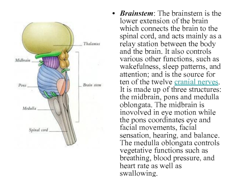
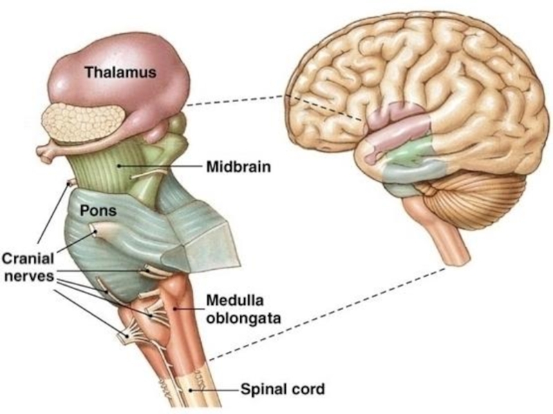
of the brain. The arachnoid layer is a thin membrane that covers the entire brain and is positioned between the dura mater and the pia mater, and for the most part does not follow the folds of the brain. The pia mater, which is attached to the surface of the entire brain, follows the folds of the brain and has many blood vessels that reach deep into the brain. The space between the arachnoid layer and the pia mater is called the subarachnoid space and it contains the cerebrospinal fluid.Слайд 10Brainstem: The brainstem is the lower extension of the brain
which connects the brain to the spinal cord, and acts
mainly as a relay station between the body and the brain. It also controls various other functions, such as wakefulness, sleep patterns, and attention; and is the source for ten of the twelve cranial nerves. It is made up of three structures: the midbrain, pons and medulla oblongata. The midbrain is inovolved in eye motion while the pons coordinates eye and facial movements, facial sensation, hearing, and balance. The medulla oblongata controls vegetative functions such as breathing, blood pressure, and heart rate as well as swallowing.Слайд 12Thalamus: The thalamus is a structure that is located above
the brainstem and it serves as a relay station for
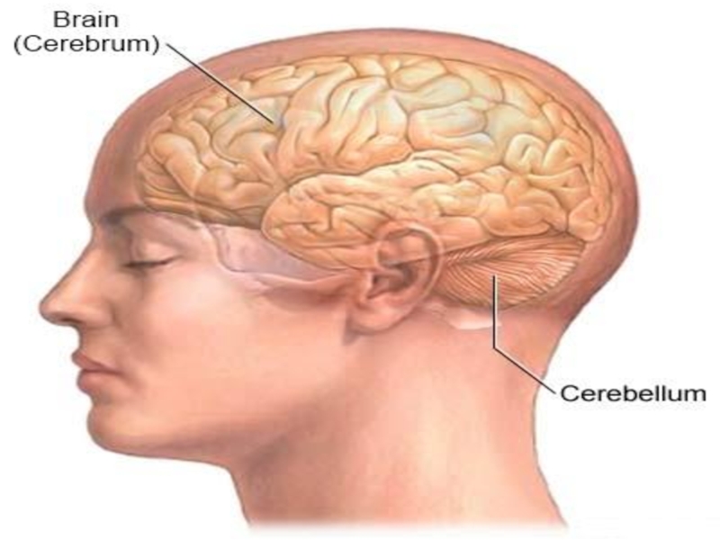
nearly all messages that travel from the cerebral cortex to the rest of the body/brain and vice versa. As such, problems within the thalamus can cause significant symptoms with regard to a variety of functions, including movement, sensation, and coordination. The thalamus also functions as an important component of the pathways within the brain that control pain sensation, attention, and wakefulness.Cerebellum: The cerebellum is located at the lower back of the brain beneath the occipital lobes and is separated from them by the tentorium. This part of the brain is responsible for maintaining balance and coordinating movements. Abnormalities in either side of the cerebellum produce symptoms on the same side of the body.
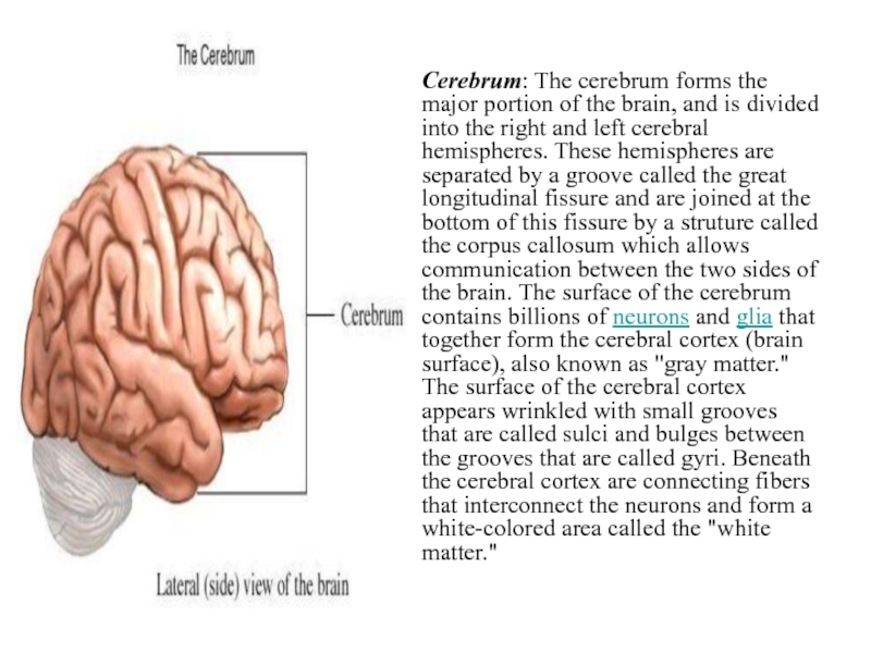
Слайд 14Cerebrum: The cerebrum forms the major portion of the brain,
and is divided into the right and left cerebral hemispheres.
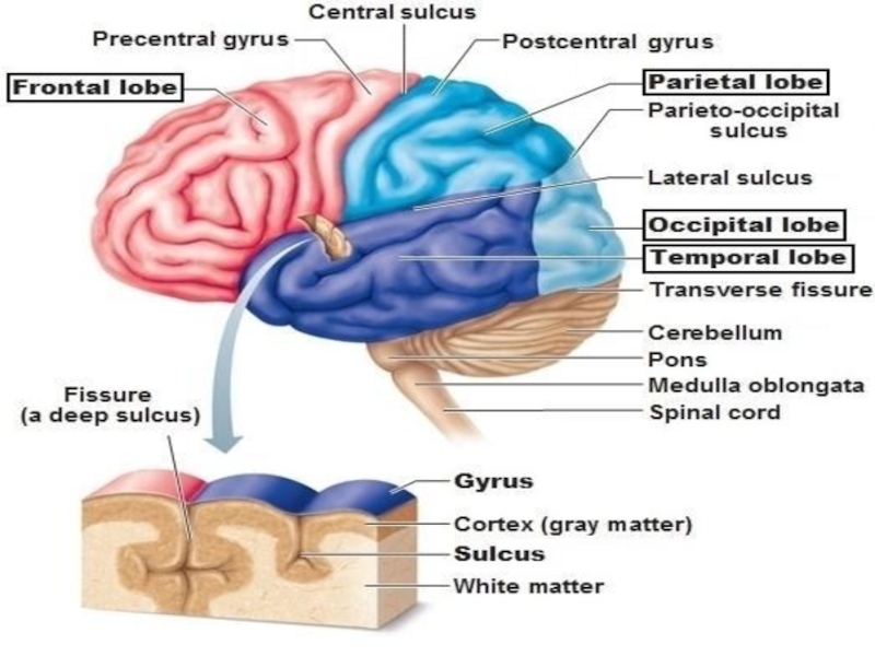
These hemispheres are separated by a groove called the great longitudinal fissure and are joined at the bottom of this fissure by a struture called the corpus callosum which allows communication between the two sides of the brain. The surface of the cerebrum contains billions of neurons and glia that together form the cerebral cortex (brain surface), also known as "gray matter." The surface of the cerebral cortex appears wrinkled with small grooves that are called sulci and bulges between the grooves that are called gyri. Beneath the cerebral cortex are connecting fibers that interconnect the neurons and form a white-colored area called the "white matter."Слайд 16Lobes: Lobes: Several large grooves (fissures) separate each side of
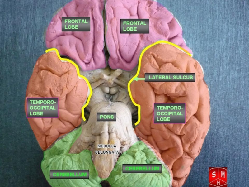
the brain into four distinct regions called lobes: frontal, temporal,
parietal, and occipital. Each hemisphere has one of each of these lobes, which generally control function on the opposite side of the body. The different portions of each lobe and the four different lobes communicate and function together through very complex relationships, but each one also has its own unique characteristics. The frontal lobes are responsible for voluntary movement, speech, intellectual and behavioral functions, memory, intelligence, concentration, temper and personality. The parietal lobe processes signals received from other areas of the brain (such as vision, hearing, motor, sensory and memory) and uses it to give meaning to objects. The occipital lobe is responsible for processing visual information. The temporal lobe is involved in visual memory and allows for recognition of objects and peoples' faces, as well as verbal memory which allows for remembering and understanding language.Слайд 18The parietal lobe processes signals received from other areas of
the brain (such as vision, hearing, motor, sensory and memory)
and uses it to give meaning to objects. The occipital lobe is responsible for processing visual information. The temporal lobe is involved in visual memory and allows for recognition of objects and peoples' faces, as well as verbal memory which allows for remembering and understanding language.Слайд 19Hypothalamus: The hypothalamus is a structure that communicates with the
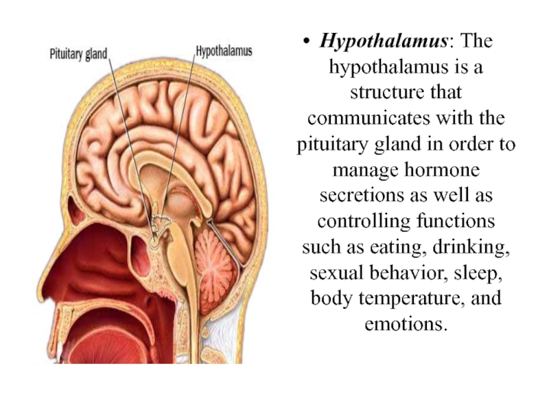
pituitary gland in order to manage hormone secretions as well
as controlling functions such as eating, drinking, sexual behavior, sleep, body temperature, and emotions.Слайд 20Pituitary Gland: The pituitary gland is a small structure that
is attached to the base of the brain in an
area called the sella turcica. This gland controls the secretion of several hormones which regulate growth and development, function of various organs (kidneys, breasts, and uterus), and the function of other glands (thyroid gland, gonads, and the adrenal glands).Basal Ganglia: The basal ganglia are clusters of nerve cells around the thalamus which are heavily connected to the cells of the cerebral cortex. The basal ganglia are associated with a variety of functions, including voluntary movement, procedural learning, eye movements, and cognitive/emotional functions. The various components of the basal ganglia include caudate nucleus, putamen, globus pallidus, substantia nigra, and subthalamic nucleus. Diseases affecting these parts can cause a number of neurological conditions, including Parkinson's disease and Huntington's disease.
