Разделы презентаций
- Разное
- Английский язык
- Астрономия
- Алгебра
- Биология
- География
- Геометрия
- Детские презентации
- Информатика
- История
- Литература
- Математика
- Медицина
- Менеджмент
- Музыка
- МХК
- Немецкий язык
- ОБЖ
- Обществознание
- Окружающий мир
- Педагогика
- Русский язык
- Технология
- Физика
- Философия
- Химия
- Шаблоны, картинки для презентаций
- Экология
- Экономика
- Юриспруденция
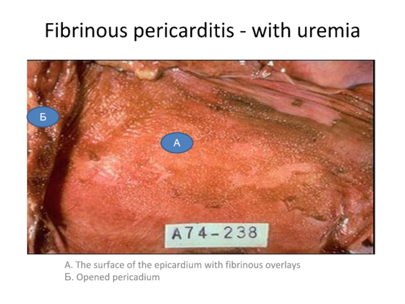
Fibrinous pericarditis - with uremia
Содержание
- 1. Fibrinous pericarditis - with uremia
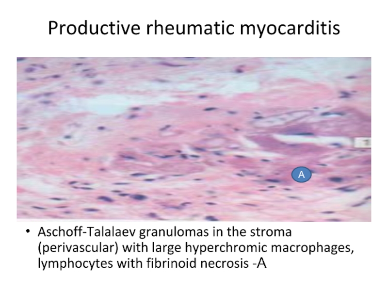
- 2. Productive rheumatic myocarditisAschoff-Talalaev granulomas in the stroma
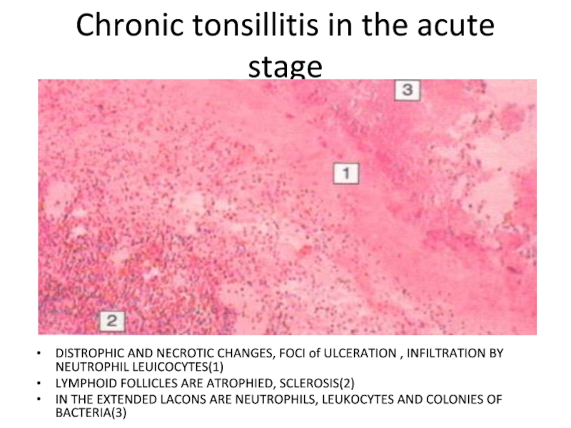
- 3. Chronic tonsillitis in the acute stageDISTROPHIC AND
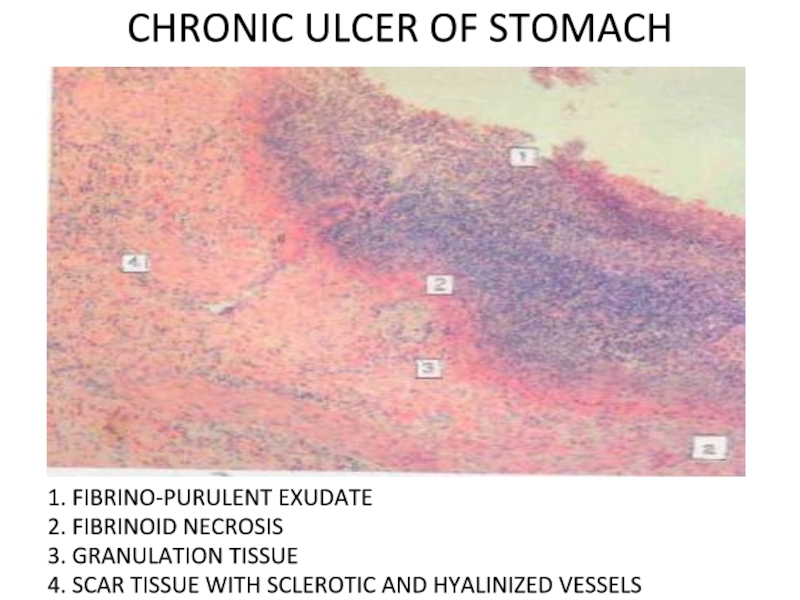
- 4. CHRONIC ULCER OF STOMACH 1. FIBRINO-PURULENT EXUDATE2.
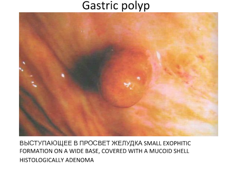
- 5. Gastric polyp ВЫСТУПАЮЩЕЕ В ПРОСВЕТ ЖЕЛУДКА SMALL
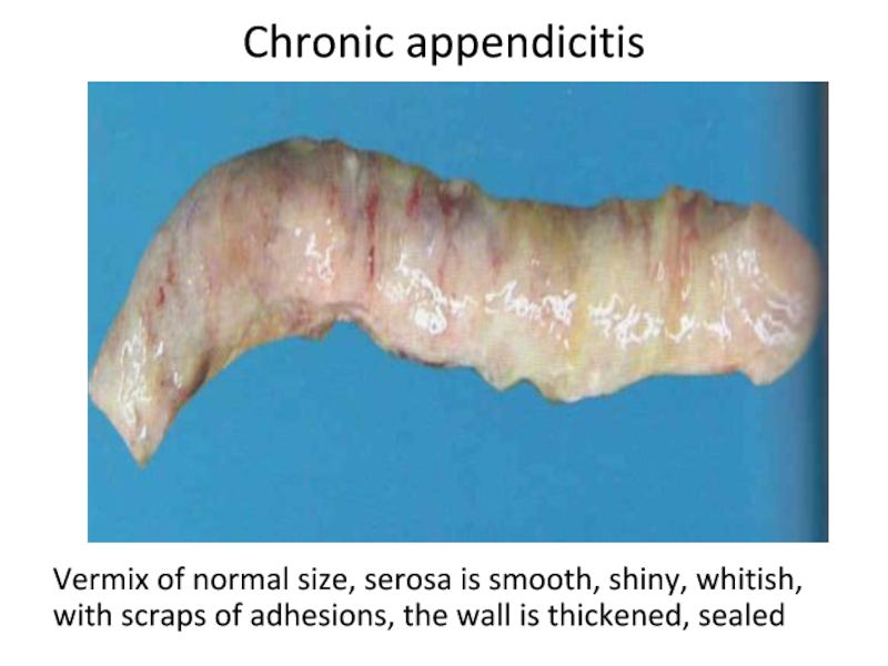
- 6. Chronic appendicitisVermix of normal size, serosa is
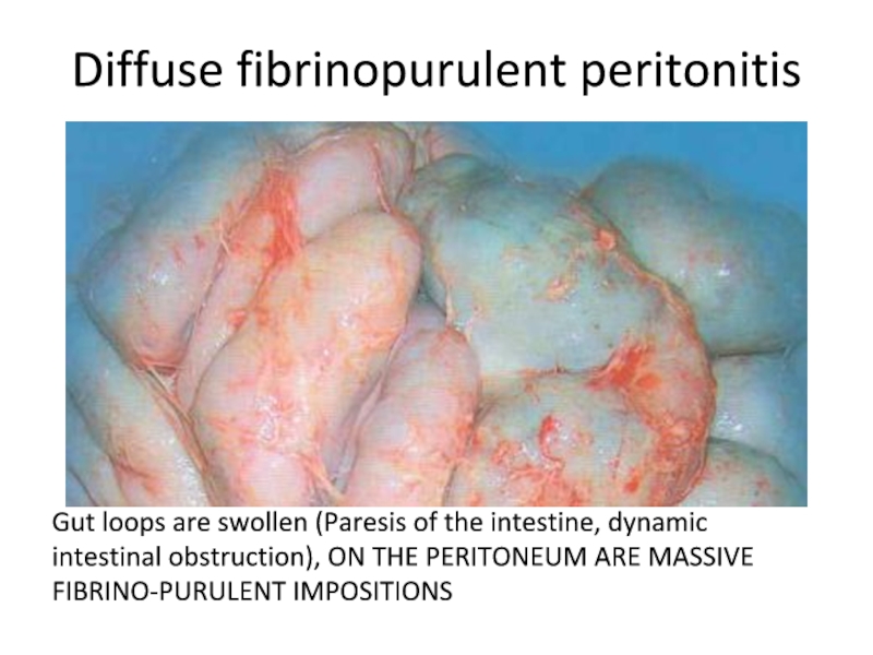
- 7. Diffuse fibrinopurulent peritonitisGut loops are swollen (Paresis
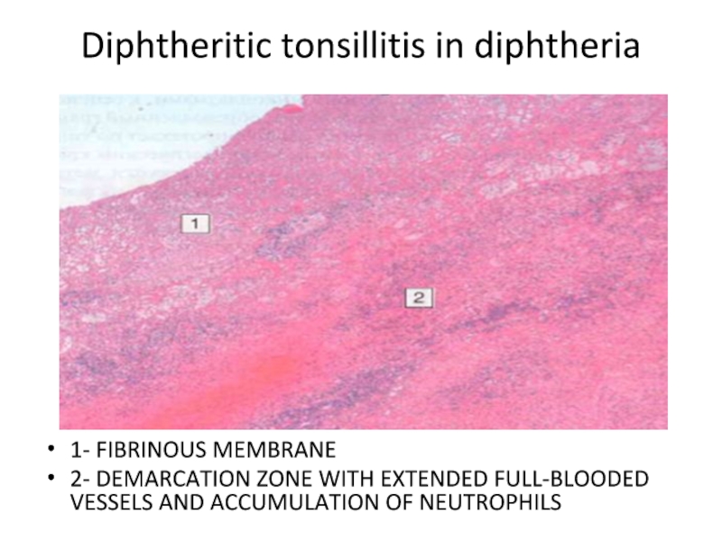
- 8. Diphtheritic tonsillitis in diphtheria 1- FIBRINOUS MEMBRANE2-
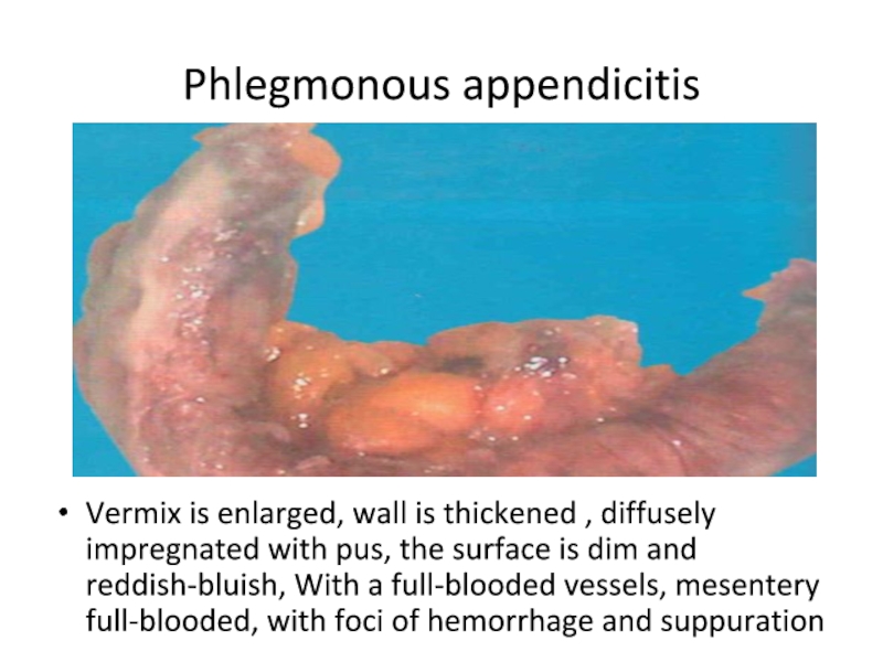
- 9. Phlegmonous appendicitis Vermix is enlarged, wall
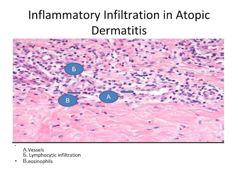
- 10. Inflammatory Infiltration in Atopic Dermatitis А.Vessels Б. Lymphocytic infiltrationВ.eosinophils ВБА
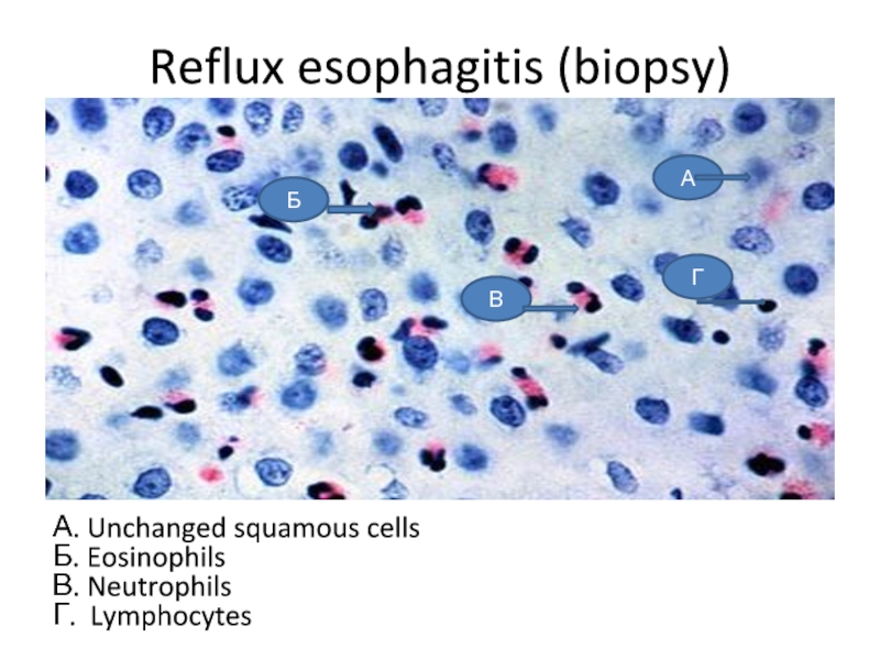
- 11. Reflux esophagitis (biopsy)А. Unchanged squamous cells Б. Eosinophils В. Neutrophils Г. Lymphocytes ГВБА
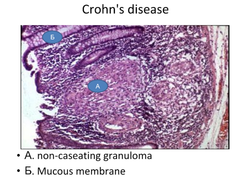
- 12. Crohn's diseaseА. non-caseating granulomaБ. Mucous membraneБА
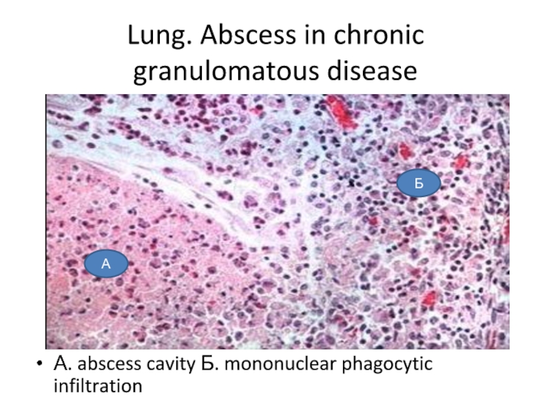
- 13. Lung. Abscess in chronic granulomatous diseaseА. abscess cavity Б. mononuclear phagocytic infiltrationАБ
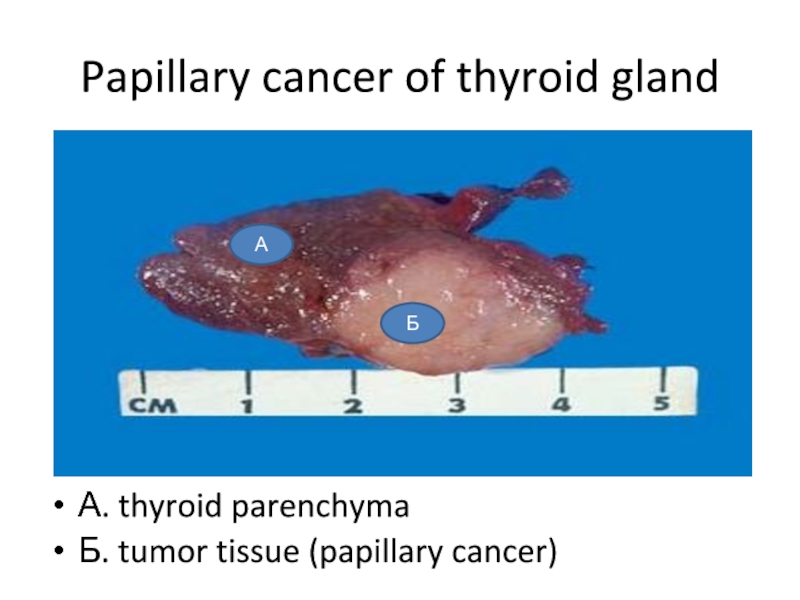
- 14. Papillary cancer of thyroid glandА. thyroid parenchymaБ. tumor tissue (papillary cancer)АБ
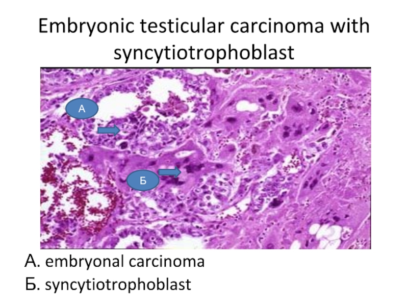
- 15. Embryonic testicular carcinoma with syncytiotrophoblastА. embryonal carcinomaБ. syncytiotrophoblastА Б
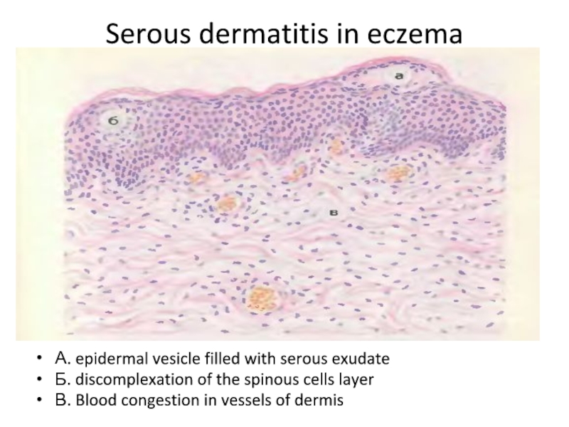
- 16. Serous dermatitis in eczemaА. epidermal vesicle filled
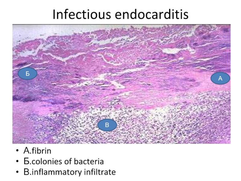
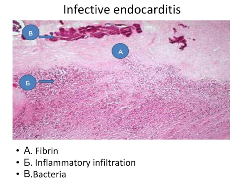
- 17. Infectious endocarditisА.fibrinБ.colonies of bacteriaВ.inflammatory infiltrateБ А В
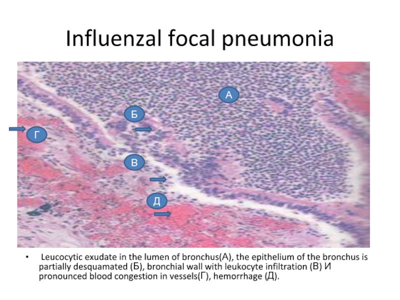
- 18. Influenzal focal pneumonia Leucocytic exudate in the
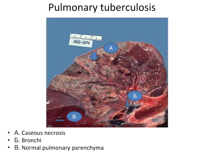
- 19. Pulmonary tuberculosis А. Caseous necrosisБ. BronchiВ. Normal pulmonary parenchymaБАВ
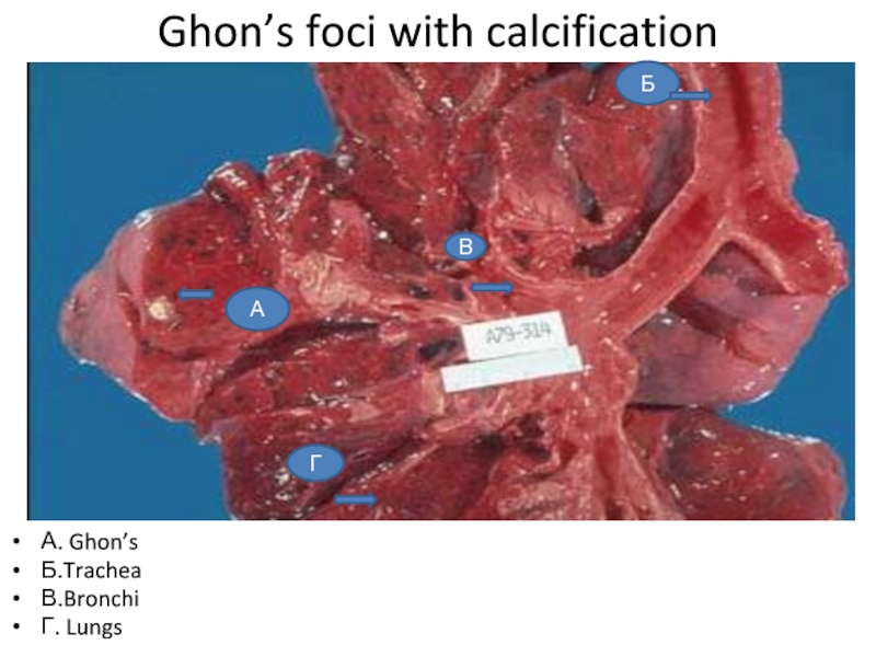
- 20. Ghon’s foci with calcification А. Ghon’s Б.TracheaВ.BronchiГ. LungsГАВБ
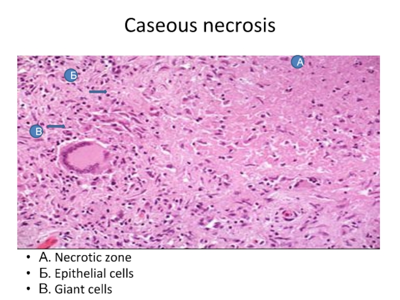
- 21. Caseous necrosis А. Necrotic zoneБ. Epithelial cellsВ. Giant cellsАВБ
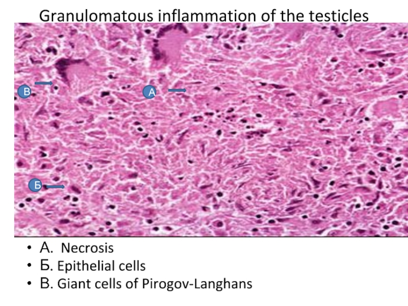
- 22. Granulomatous inflammation of the testicles А. NecrosisБ. Epithelial cellsВ. Giant cells of Pirogov-LanghansВБА
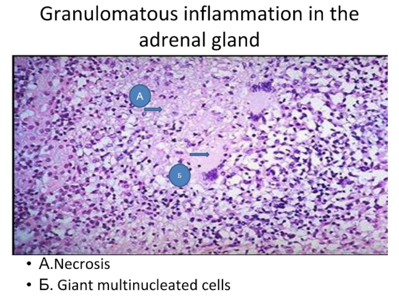
- 23. Granulomatous inflammation in the adrenal glandА.NecrosisБ. Giant multinucleated cellsА Б
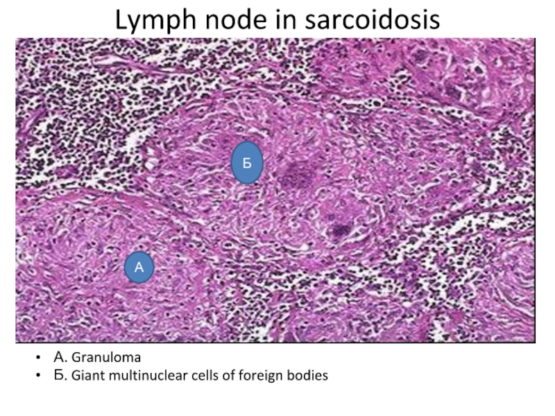
- 24. Lymph node in sarcoidosis А. GranulomaБ. Giant multinuclear cells of foreign bodiesА Б
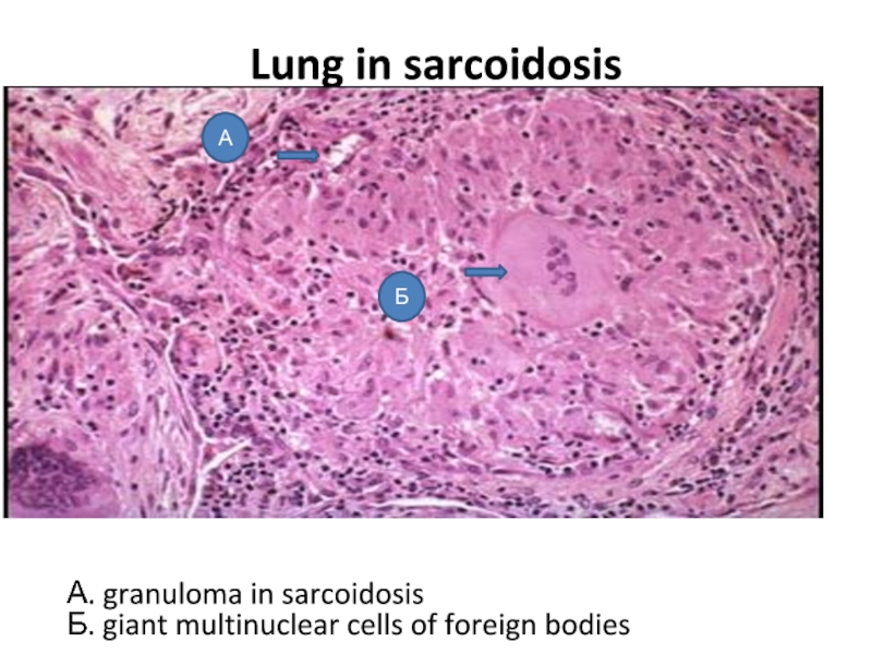
- 25. Lung in sarcoidosisБ А А. granuloma in sarcoidosisБ. giant multinuclear cells of foreign bodies
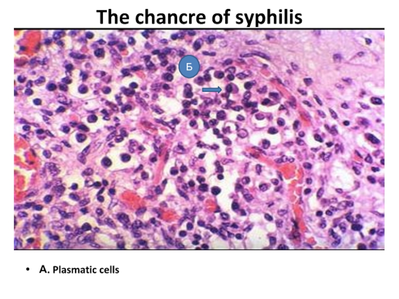
- 26. The chancre of syphilis А. Plasmatic cellsБ
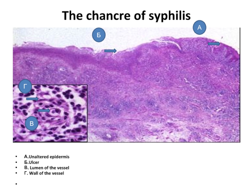
- 27. The chancre of syphilis А.Unaltered epidermisБ.UlcerВ.
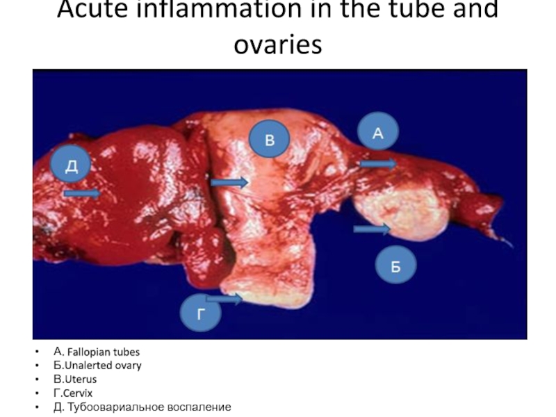
- 28. Acute inflammation in the tube and ovaries А. Fallopian tubesБ.Unalerted ovaryВ.UterusГ.CervixД. Тубоовариальное воспаление
- 29. Acute inflammation of the fallopian tubeА. Muscular
- 30. Inflammation of the pelvic organs. Fallopian tube А.Lumen of the vesselБ. Mucous membraneВ. SubmucosaГ.Plasmatic cellsД.Neutrophils
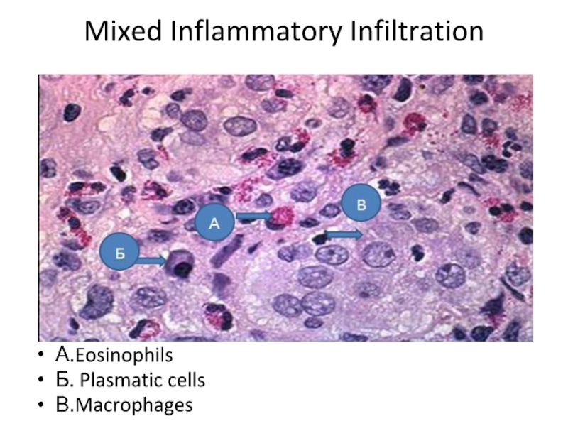
- 31. Mixed Inflammatory Infiltration А.EosinophilsБ. Plasmatic cellsВ.Macrophages
- 32. Pulmonary tuberculosis А. caseous necrosisБ. shaft of epithelioid cellsВ. giant cells of Pirogov-Langhans
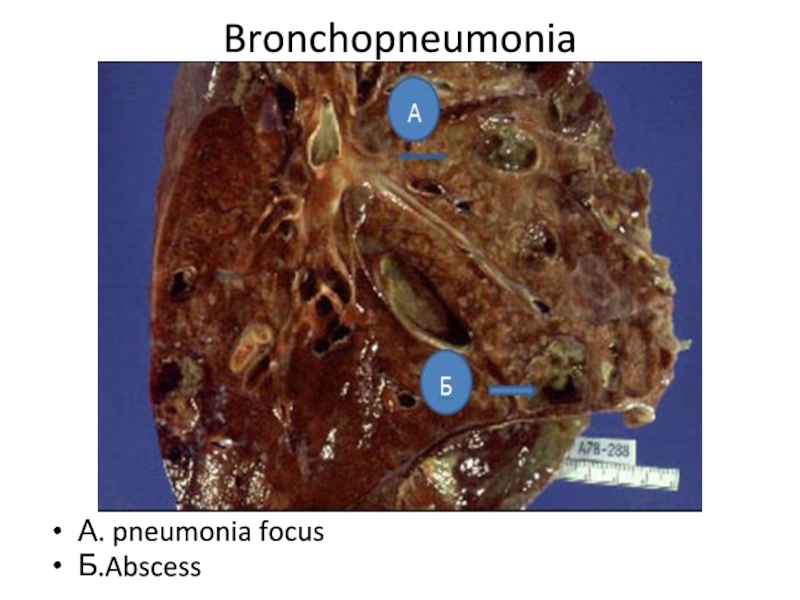
- 33. BronchopneumoniaА. pneumonia focusБ.Abscess
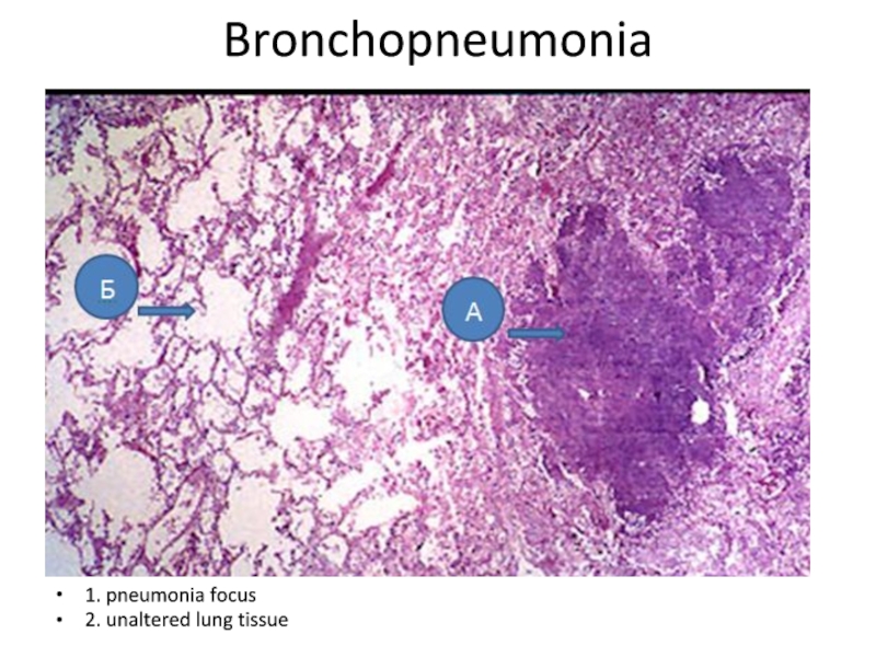
- 34. Bronchopneumonia1. pneumonia focus2. unaltered lung tissue
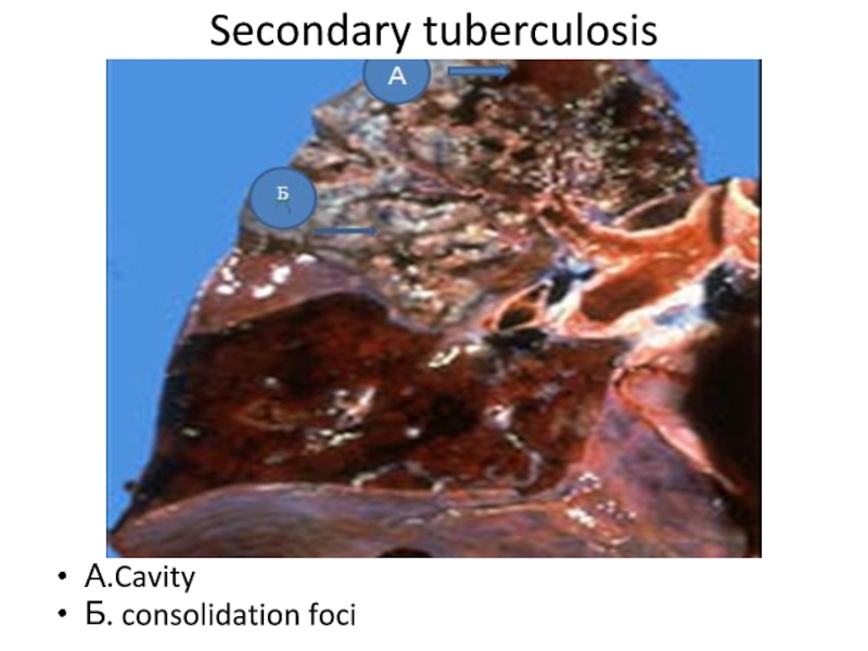
- 35. Secondary tuberculosis А.CavityБ. consolidation foci
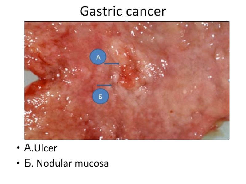
- 36. Gastric cancerА.UlcerБ. Nodular mucosa
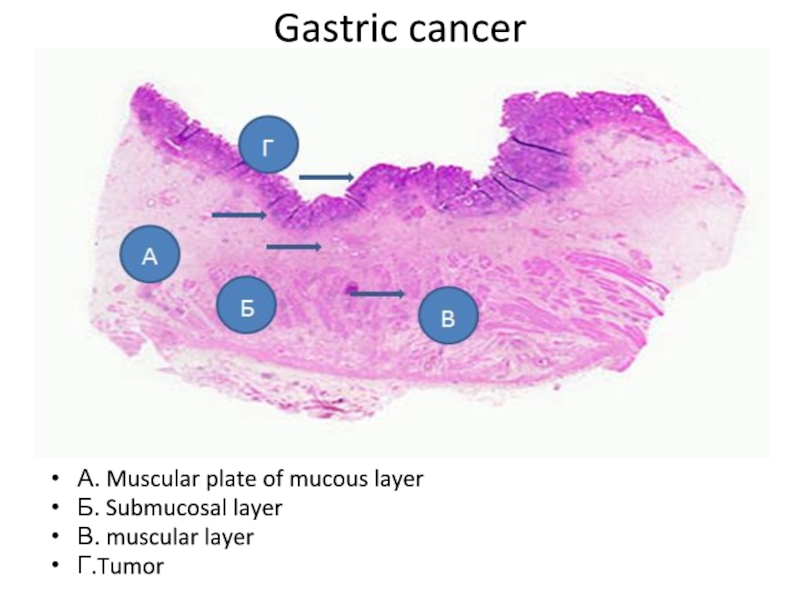
- 37. Gastric cancer А. Muscular plate of mucous layerБ. Submucosal layerВ. muscular layerГ.Tumor
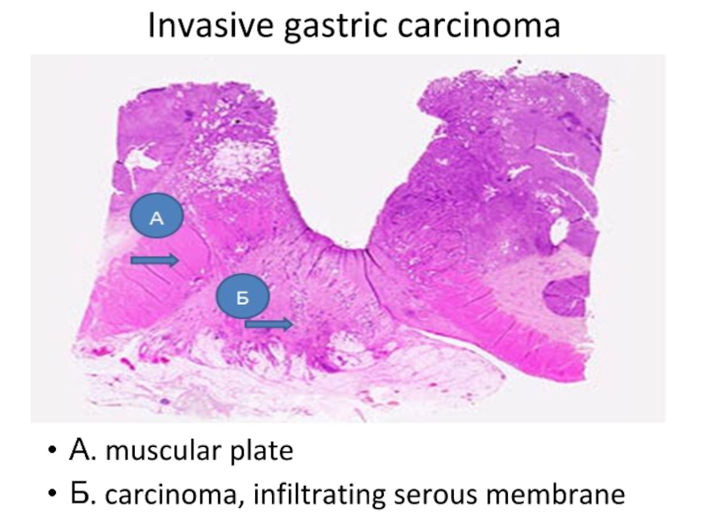
- 38. Invasive gastric carcinoma А. muscular plateБ. carcinoma, infiltrating serous membrane
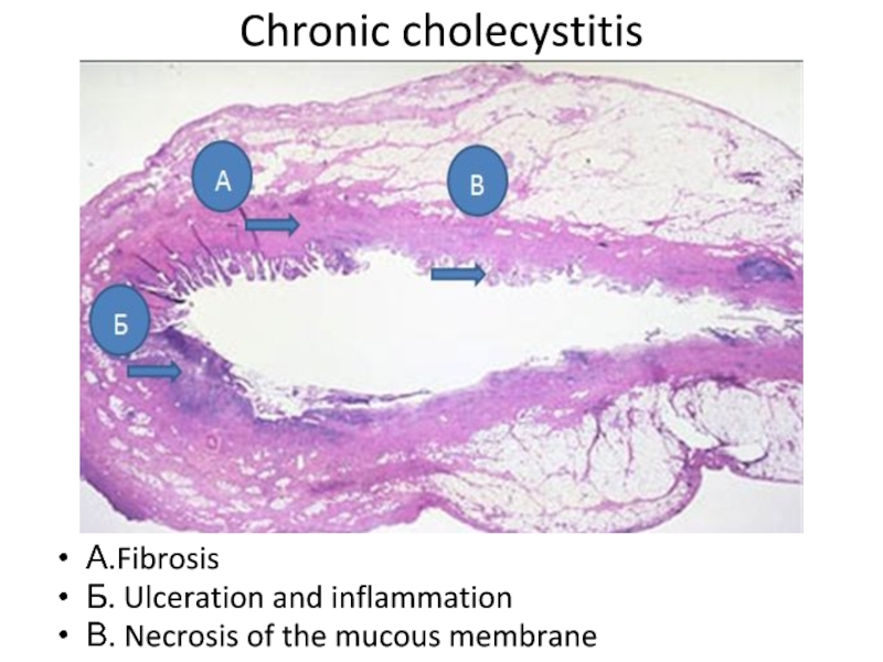
- 39. Chronic cholecystitis А.FibrosisБ. Ulceration and inflammationВ. Necrosis of the mucous membrane
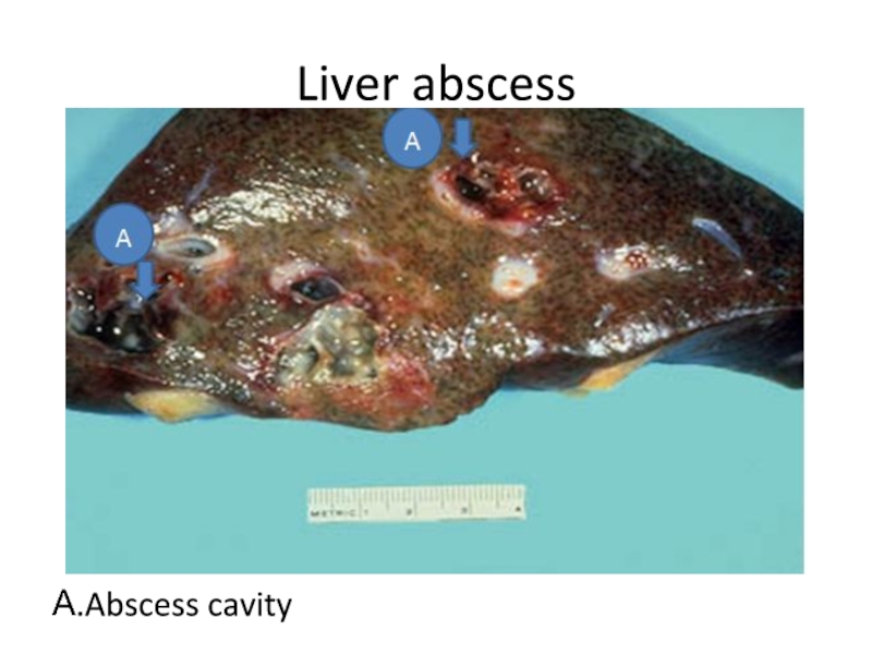
- 40. Liver abscessА.Abscess cavity
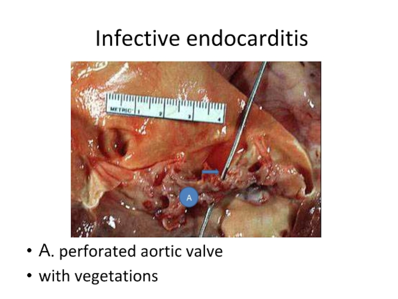
- 41. Infective endocarditisА. perforated aortic valvewith vegetations
- 42. Infective endocarditis А. FibrinБ. Inflammatory infiltrationВ.Bacteria
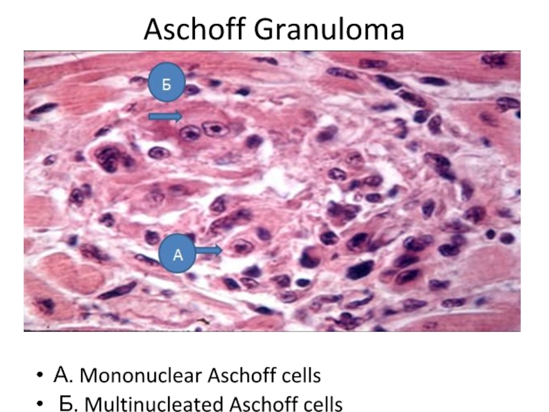
- 43. Aschoff GranulomaА. Mononuclear Aschoff cells Б. Multinucleated Aschoff cells
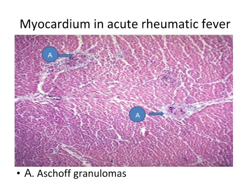
- 44. Myocardium in acute rheumatic feverА. Aschoff granulomas
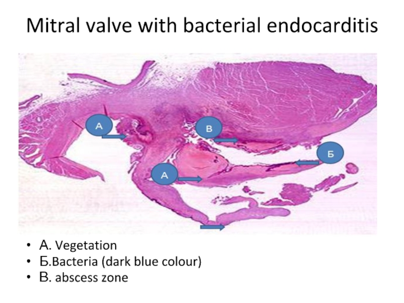
- 45. Mitral valve with bacterial endocarditis А. VegetationБ.Bacteria (dark blue colour)В. abscess zone
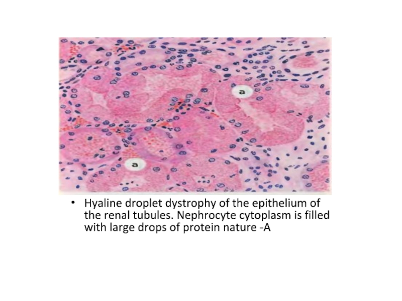
- 46. Hyaline droplet dystrophy of the epithelium of
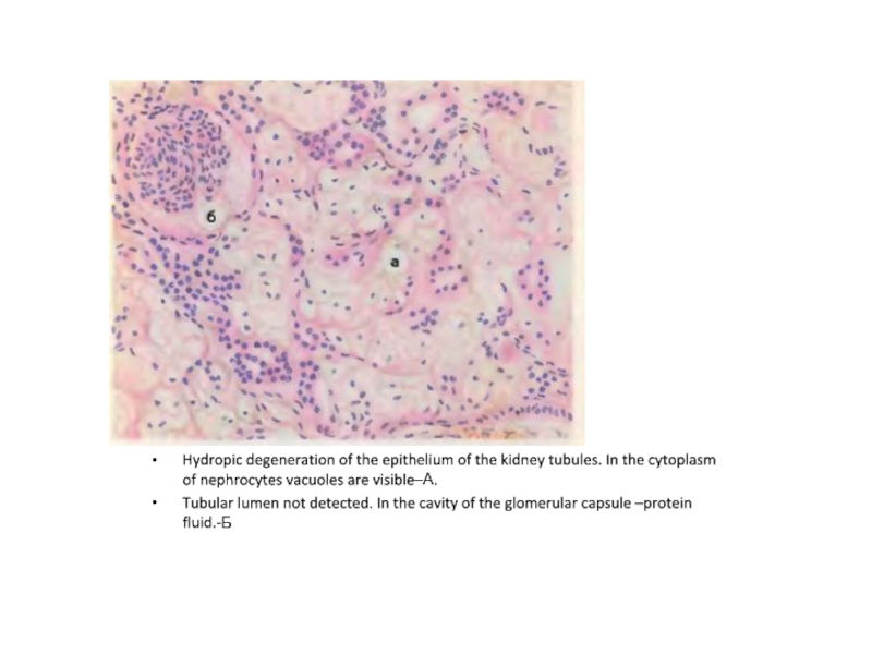
- 47. Hydropic degeneration of the epithelium of the
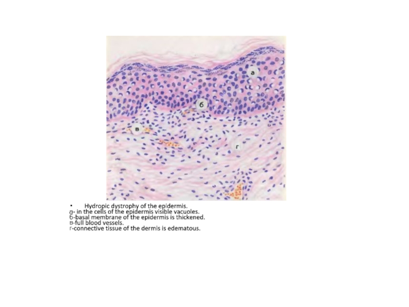
- 48. Hydropic dystrophy of the epidermis. а- in
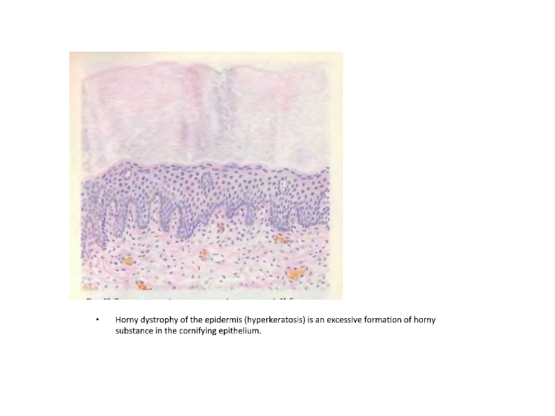
- 49. Horny dystrophy of the epidermis (hyperkeratosis) is
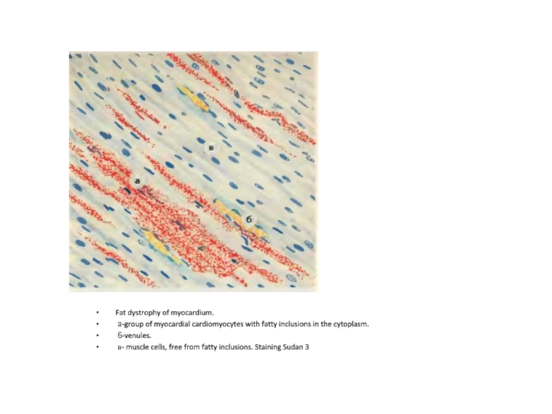
- 50. Fat dystrophy of myocardium. а-group of myocardial
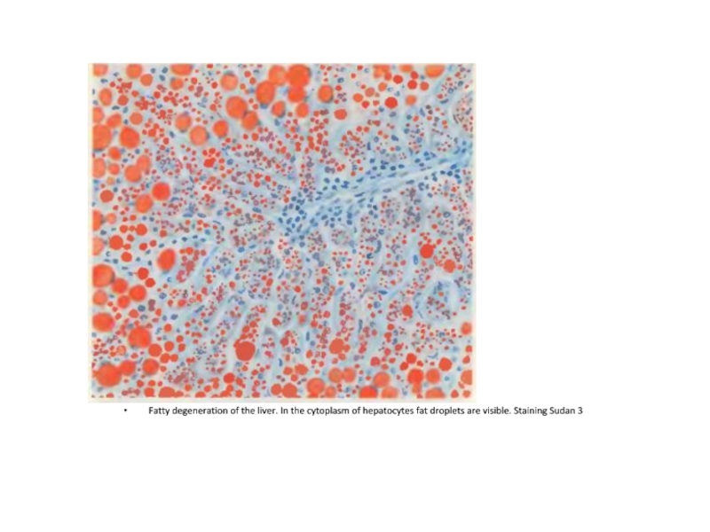
- 51. Fatty degeneration of the liver. In the
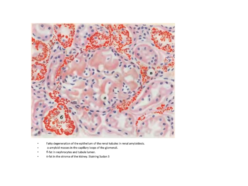
- 52. Fatty degeneration of the epithelium of the
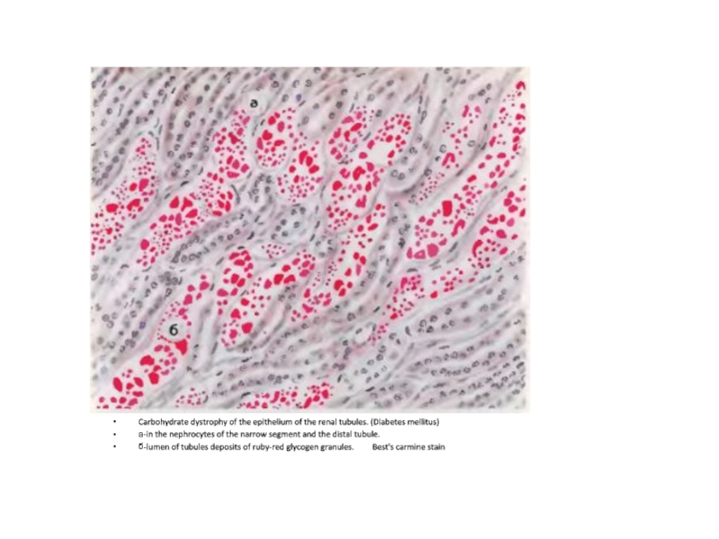
- 53. Carbohydrate dystrophy of the epithelium of the
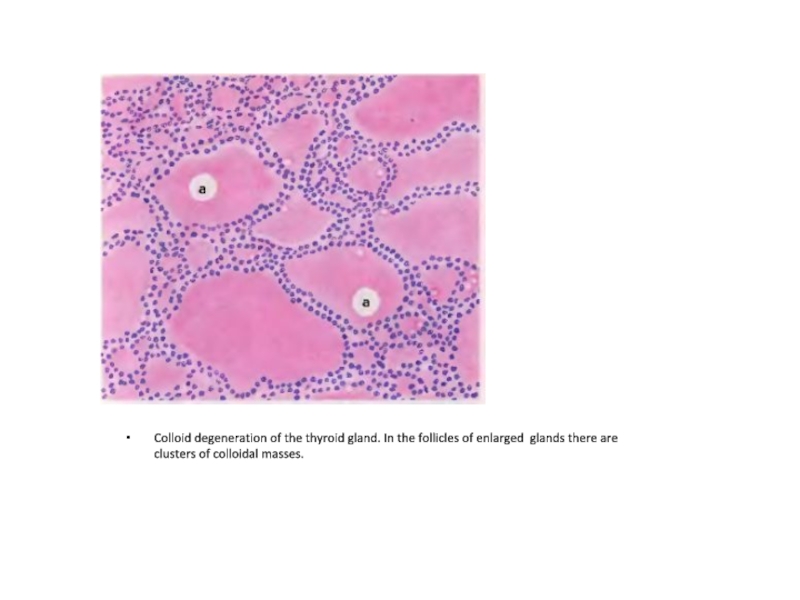
- 54. Colloid degeneration of the thyroid gland. In
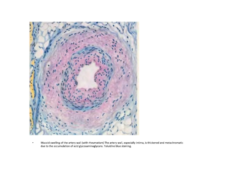
- 55. Mucoid swelling of the artery wall (with
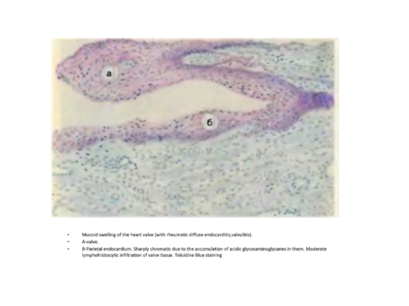
- 56. Mucoid swelling of the heart valve (with
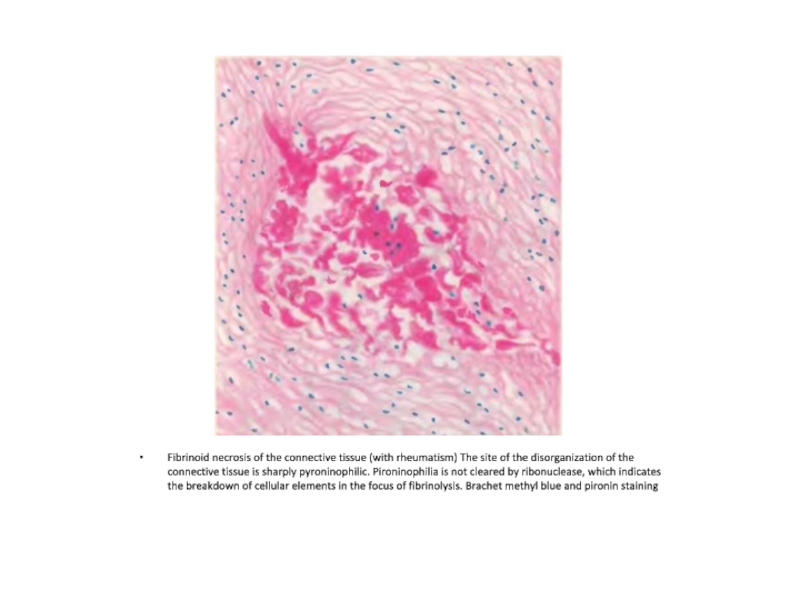
- 57. Fibrinoid necrosis of the connective tissue (with
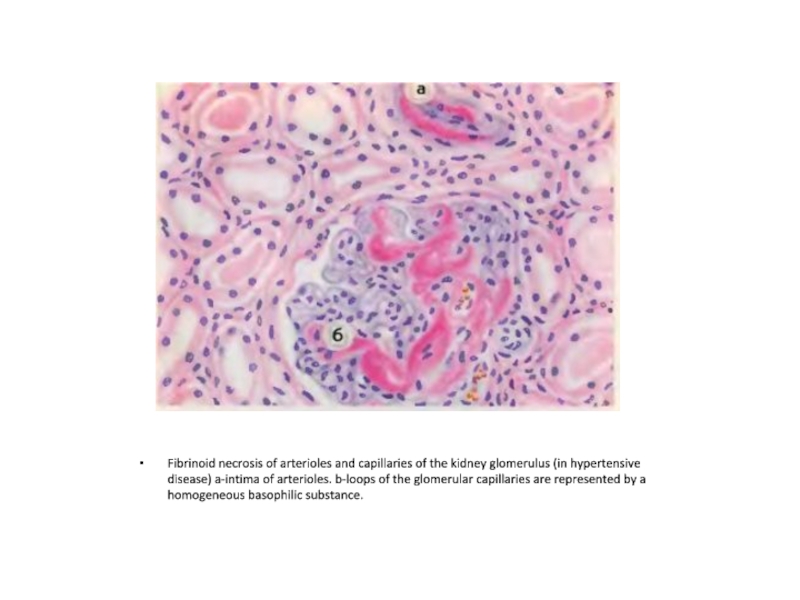
- 58. Fibrinoid necrosis of arterioles and capillaries of
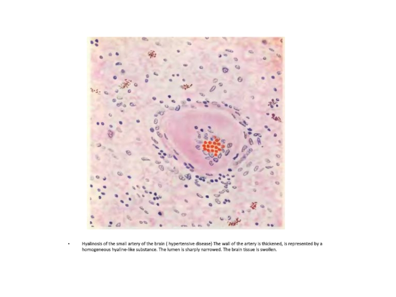
- 59. Hyalinosis of the small artery of the
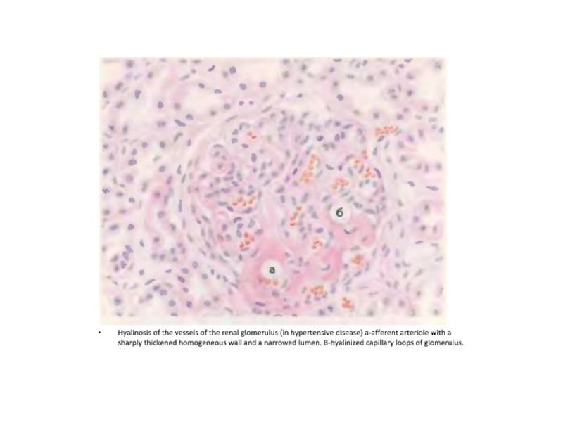
- 60. Hyalinosis of the vessels of the renal
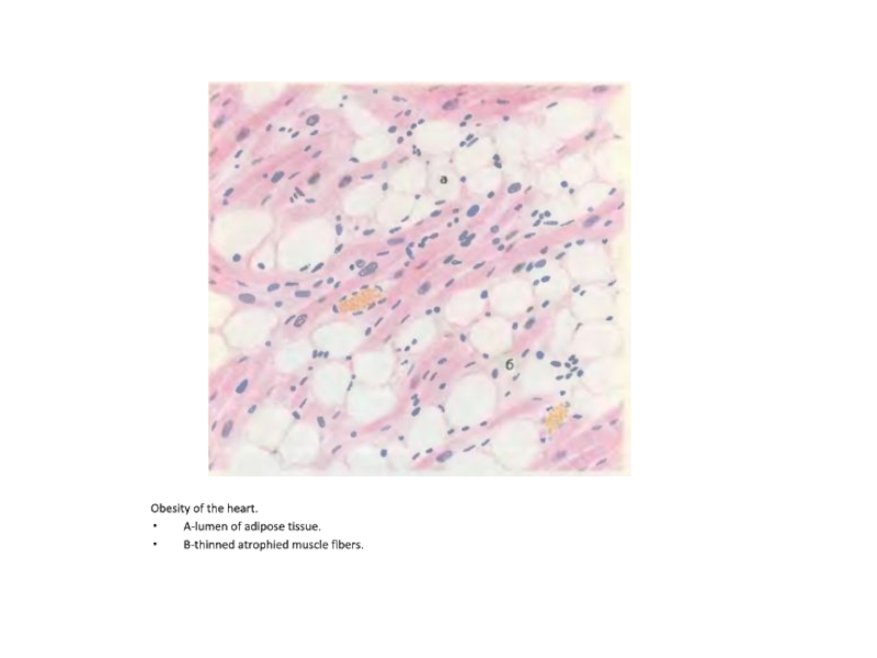
- 61. Obesity of the heart. A-lumen of adipose tissue. B-thinned atrophied muscle fibers.
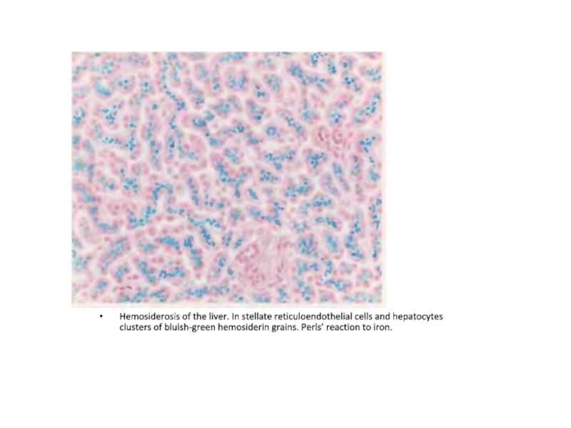
- 62. Hemosiderosis of the liver. In stellate reticuloendothelial
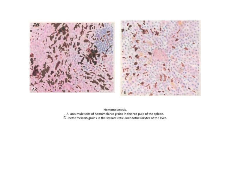
- 63. Hemomelanosis. A- accumulations of hemomelanin grains
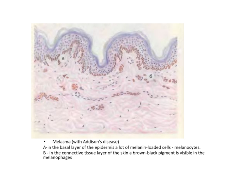
- 64. Melasma (with Addison's disease) A-in the basal
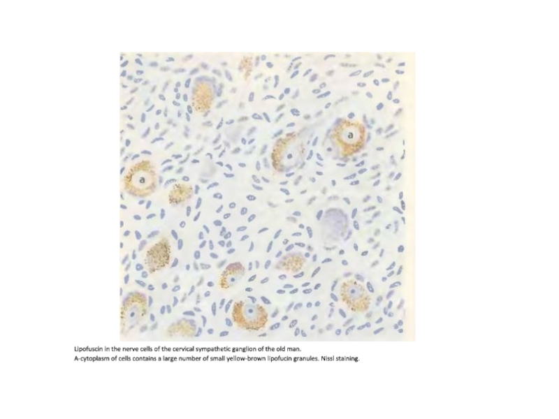
- 65. Lipofuscin in the nerve cells of the
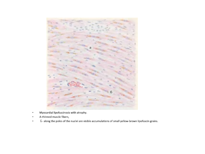
- 66. Myocardial lipofuscinosis with atrophy. A-thinned muscle fibers,
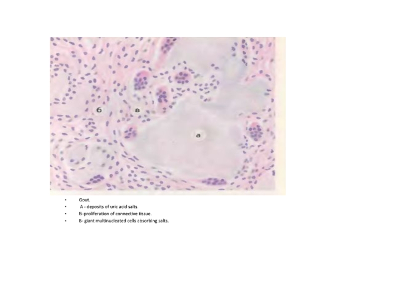
- 67. Gout. A - deposits of uric acid
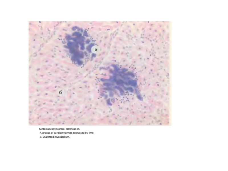
- 68. Metastatic myocardial calcification. A-groups of cardiomyocytes encrusted by lime. Б-unalerted myocardium.
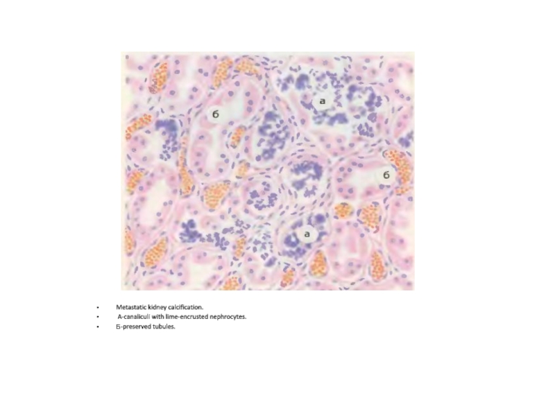
- 69. Metastatic kidney calcification. A-canaliculi with lime-encrusted nephrocytes. Б-preserved tubules.
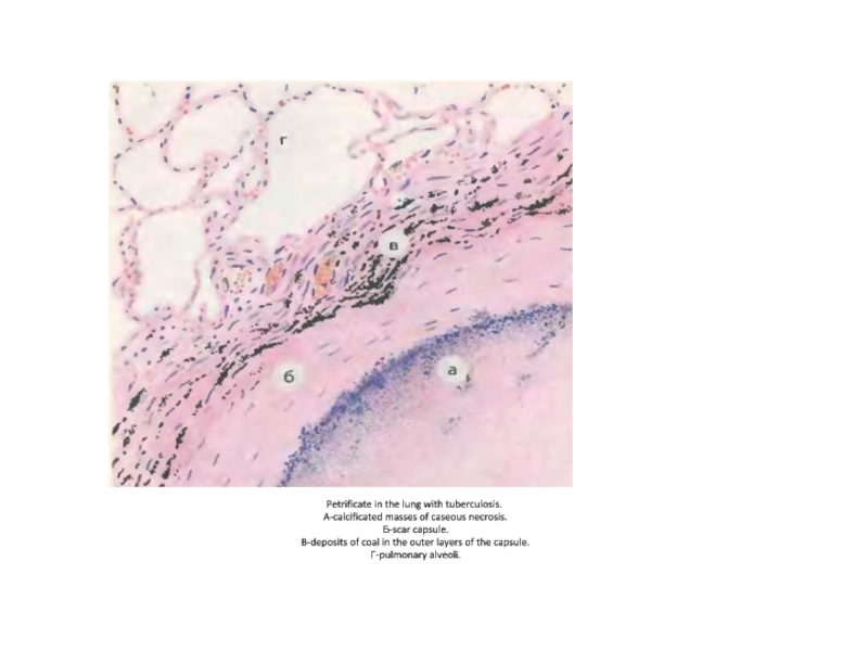
- 70. Petrificate in the lung with tuberculosis.
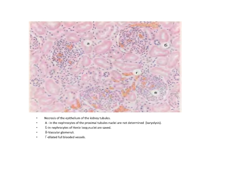
- 71. Necrosis of the epithelium of the kidney
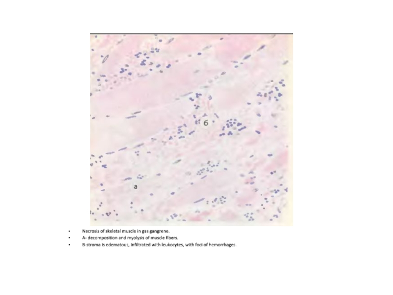
- 72. Necrosis of skeletal muscle in gas gangrene.
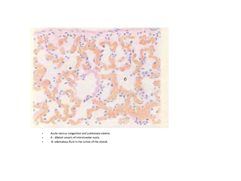
- 73. Acute venous congestion and pulmonary edema. A
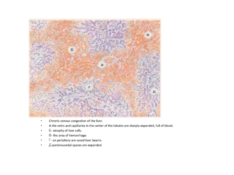
- 74. Chronic venous congestion of the liver. A-the
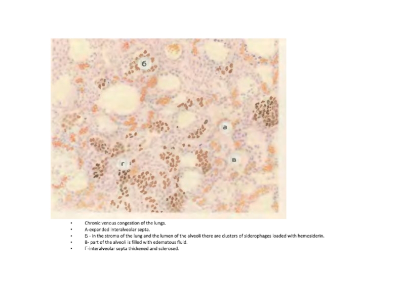
- 75. Chronic venous congestion of the lungs. A-expanded
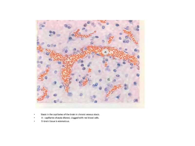
- 76. Stasis in the capillaries of the brain
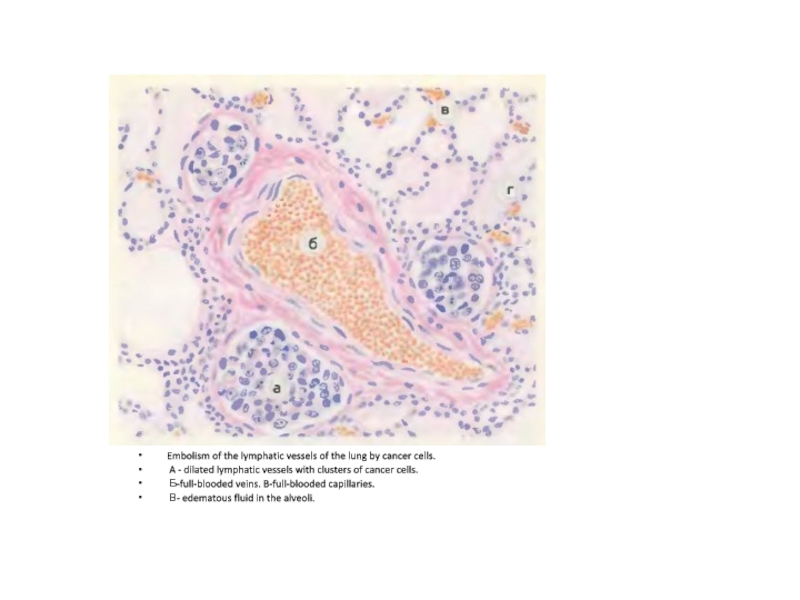
- 77. Embolism of the lymphatic vessels of the
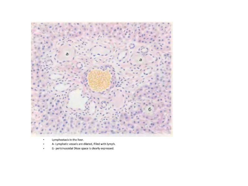
- 78. Lymphostasis in the liver. A- Lymphatic vessels
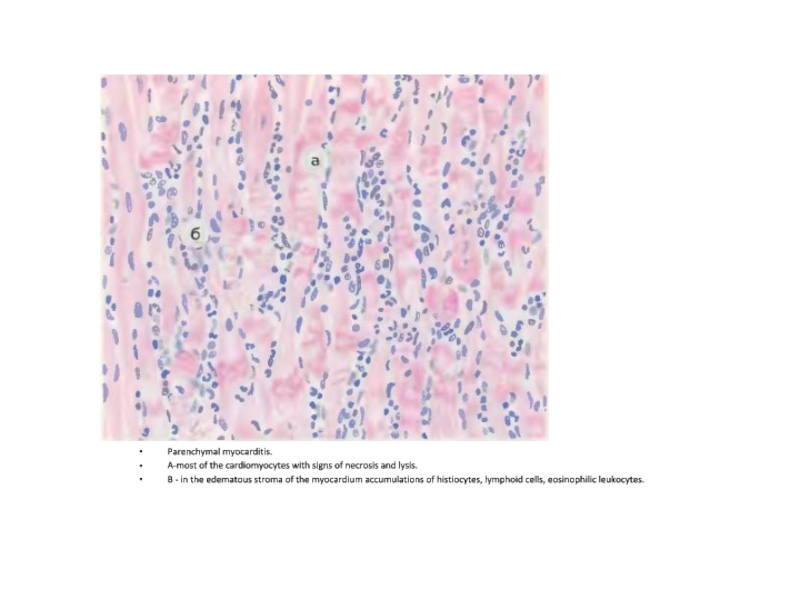
- 79. Parenchymal myocarditis. A-most of the cardiomyocytes with
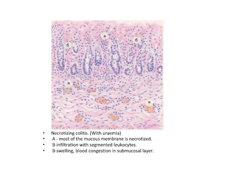
- 80. Necrotizing colitis. (With uraemia) A - most
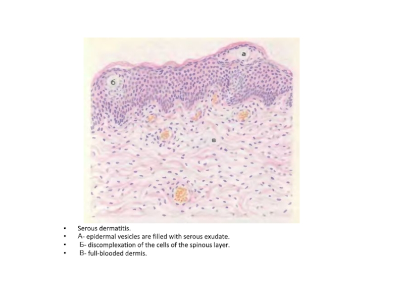
- 81. Serous dermatitis. А- epidermal vesicles are filled
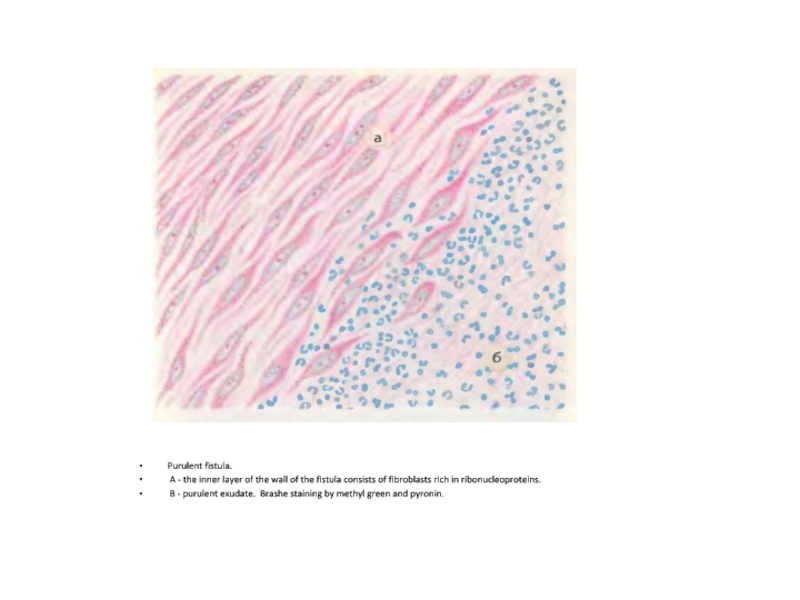
- 82. Purulent fistula. A - the inner layer
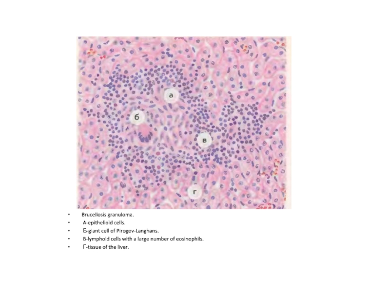
- 83. Brucellosis granuloma. A-epithelioid cells. Б-giant cell of
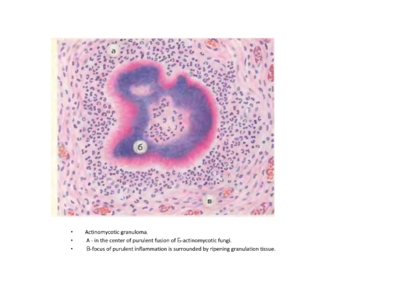
- 84. Actinomycotic granuloma. A - in the center
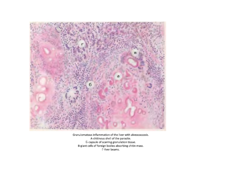
- 85. Granulomatous inflammation of the liver with alveococcosis.
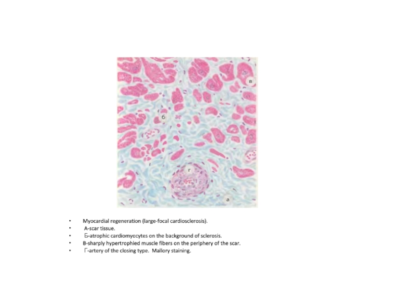
- 86. Myocardial regeneration (large-focal cardiosclerosis). A-scar tissue. Б-atrophic
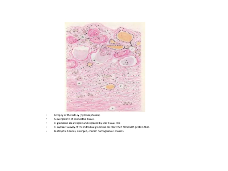
- 87. Atrophy of the kidney (hydronephrosis). A-overgrowth of
- 88. Скачать презентанцию
Productive rheumatic myocarditisAschoff-Talalaev granulomas in the stroma (perivascular) with large hyperchromic macrophages, lymphocytes with fibrinoid necrosis -АА
Слайды и текст этой презентации
Слайд 1Fibrinous pericarditis - with uremia
A. The surface of the epicardium
with fibrinous overlays
Слайд 2Productive rheumatic myocarditis
Aschoff-Talalaev granulomas in the stroma (perivascular) with large
hyperchromic macrophages, lymphocytes with fibrinoid necrosis -А
А
Слайд 3Chronic tonsillitis in the acute stage
DISTROPHIC AND NECROTIC CHANGES, FOCI
of ULCERATION , INFILTRATION BY NEUTROPHIL LEUICOCYTES(1)
LYMPHOID FOLLICLES ARE ATROPHIED,
SCLEROSIS(2)IN THE EXTENDED LACONS ARE NEUTROPHILS, LEUKOCYTES AND COLONIES OF BACTERIA(3)
Слайд 4CHRONIC ULCER OF STOMACH
1. FIBRINO-PURULENT EXUDATE
2. FIBRINOID NECROSIS
3. GRANULATION TISSUE
4.
SCAR TISSUE WITH SCLEROTIC AND HYALINIZED VESSELS
Слайд 5Gastric polyp
ВЫСТУПАЮЩЕЕ В ПРОСВЕТ ЖЕЛУДКА SMALL EXOPHITIC FORMATION ON A
WIDE BASE, COVERED WITH A MUCOID SHELL
HISTOLOGICALLY ADENOMA
Слайд 6Chronic appendicitis
Vermix of normal size, serosa is smooth, shiny, whitish,
with scraps of adhesions, the wall is thickened, sealed
Слайд 7Diffuse fibrinopurulent peritonitis
Gut loops are swollen (Paresis of the intestine,
dynamic intestinal obstruction), ON THE PERITONEUM ARE MASSIVE FIBRINO-PURULENT IMPOSITIONS
Слайд 8Diphtheritic tonsillitis in diphtheria
1- FIBRINOUS MEMBRANE
2- DEMARCATION ZONE WITH EXTENDED
FULL-BLOODED VESSELS AND ACCUMULATION OF NEUTROPHILS
Слайд 9
Phlegmonous appendicitis
Vermix is enlarged, wall is thickened , diffusely impregnated
with pus, the surface is dim and reddish-bluish, With a
full-blooded vessels, mesentery full-blooded, with foci of hemorrhage and suppurationСлайд 10Inflammatory Infiltration in Atopic Dermatitis
А.Vessels
Б. Lymphocytic infiltration
В.eosinophils
В
Б
А
Слайд 11Reflux esophagitis (biopsy)
А. Unchanged squamous cells
Б. Eosinophils
В. Neutrophils
Г. Lymphocytes
Г
В
Б
А
Слайд 13Lung. Abscess in chronic granulomatous disease
А. abscess cavity Б. mononuclear
phagocytic infiltration
А
Б
Слайд 15Embryonic testicular carcinoma with syncytiotrophoblast
А. embryonal carcinoma
Б. syncytiotrophoblast
А
Б
Слайд 16Serous dermatitis in eczema
А. epidermal vesicle filled with serous exudate
Б.
discomplexation of the spinous cells layer
В. Blood congestion in vessels
of dermisСлайд 18Influenzal focal pneumonia
Leucocytic exudate in the lumen of bronchus(А),
the epithelium of the bronchus is partially desquamated (Б), bronchial
wall with leukocyte infiltration (В) И pronounced blood congestion in vessels(Г), hemorrhage (Д).В
Б
А
Д
Г
Слайд 22Granulomatous inflammation of the testicles
А. Necrosis
Б. Epithelial cells
В. Giant cells
of Pirogov-Langhans
В
Б
А
Слайд 25
Lung in sarcoidosis
Б
А
А. granuloma in sarcoidosis
Б. giant multinuclear
cells of foreign bodies
Слайд 27
The chancre of syphilis
А.Unaltered epidermis
Б.Ulcer
В. Lumen of the vessel
Г. Wall
of the vessel
Б
А
Г
В
Слайд 28Acute inflammation in the tube and ovaries
А. Fallopian tubes
Б.Unalerted ovary
В.Uterus
Г.Cervix
Д.
Тубоовариальное воспаление
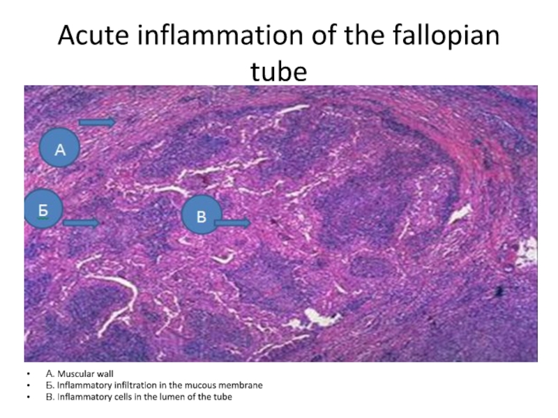
Слайд 29Acute inflammation of the fallopian tube
А. Muscular wall
Б. Inflammatory infiltration
in the mucous membrane
В. Inflammatory cells in the lumen of
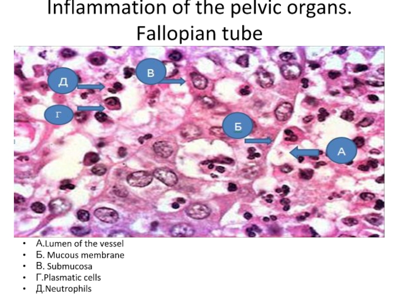
the tubeСлайд 30Inflammation of the pelvic organs. Fallopian tube
А.Lumen of the vessel
Б.
Mucous membrane
В. Submucosa
Г.Plasmatic cells
Д.Neutrophils
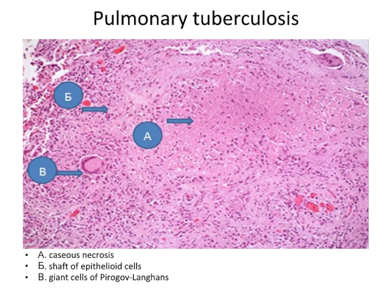
Слайд 32Pulmonary tuberculosis
А. caseous necrosis
Б. shaft of epithelioid cells
В. giant cells
of Pirogov-Langhans
Слайд 39
Chronic cholecystitis
А.Fibrosis
Б. Ulceration and inflammation
В. Necrosis of the mucous membrane
Слайд 45Mitral valve with bacterial endocarditis
А. Vegetation
Б.Bacteria (dark blue colour)
В. abscess
zone
Слайд 46Hyaline droplet dystrophy of the epithelium of the renal tubules.
Nephrocyte cytoplasm is filled with large drops of protein nature
-AСлайд 47Hydropic degeneration of the epithelium of the kidney tubules. In
the cytoplasm of nephrocytes vacuoles are visible–А.
Tubular lumen not
detected. In the cavity of the glomerular capsule –protein fluid.-Б Слайд 48Hydropic dystrophy of the epidermis.
а- in the cells of
the epidermis visible vacuoles.
б-basal membrane of the epidermis is
thickened.в-full blood vessels.
г-connective tissue of the dermis is edematous.
Слайд 49Horny dystrophy of the epidermis (hyperkeratosis) is an excessive formation
of horny substance in the cornifying epithelium.
Слайд 50Fat dystrophy of myocardium.
а-group of myocardial cardiomyocytes with fatty
inclusions in the cytoplasm.
б-venules.
в- muscle cells, free from
fatty inclusions. Staining Sudan 3Слайд 51Fatty degeneration of the liver. In the cytoplasm of hepatocytes
fat droplets are visible. Staining Sudan 3
Слайд 52Fatty degeneration of the epithelium of the renal tubules in
renal amyloidosis.
а-amyloid masses in the capillary loops of the
glomeruli. б-fat in nephrocytes and tubule lumen.
в-fat in the stroma of the kidney. Staining Sudan 3
Слайд 53Carbohydrate dystrophy of the epithelium of the renal tubules. (Diabetes
mellitus)
а-in the nephrocytes of the narrow segment and the
distal tubule. б-lumen of tubules deposits of ruby-red glycogen granules. Best's carmine stain
Слайд 54Colloid degeneration of the thyroid gland. In the follicles of
enlarged glands there are clusters of colloidal masses.
Слайд 55Mucoid swelling of the artery wall (with rheumatism) The artery
wall, especially intima, is thickened and metachromatic due to the
accumulation of acid glycosaminoglycans. Toluidine blue staining.Слайд 56Mucoid swelling of the heart valve (with rheumatic diffuse endocarditis,valvulitis).
A-valve.
B-Parietal endocardium. Sharply chromatic due to the accumulation of
acidic glycosaminoglycanes in them. Moderate lymphohistiocytic infiltration of valve tissue. Toluidine Blue stainingСлайд 57Fibrinoid necrosis of the connective tissue (with rheumatism) The site
of the disorganization of the connective tissue is sharply pyroninophilic.
Pironinophilia is not cleared by ribonuclease, which indicates the breakdown of cellular elements in the focus of fibrinolysis. Brachet methyl blue and pironin stainingСлайд 58Fibrinoid necrosis of arterioles and capillaries of the kidney glomerulus
(in hypertensive disease) a-intima of arterioles. b-loops of the glomerular
capillaries are represented by a homogeneous basophilic substance.Слайд 59Hyalinosis of the small artery of the brain ( hypertensive
disease) The wall of the artery is thickened, is represented
by a homogeneous hyaline-like substance. The lumen is sharply narrowed. The brain tissue is swollen.Слайд 60Hyalinosis of the vessels of the renal glomerulus (in hypertensive
disease) a-afferent arteriole with a sharply thickened homogeneous wall and
a narrowed lumen. B-hyalinized capillary loops of glomerulus.Слайд 62Hemosiderosis of the liver. In stellate reticuloendothelial cells and hepatocytes
clusters of bluish-green hemosiderin grains. Perls’ reaction to iron.
Слайд 63Hemomelanosis. A- accumulations of hemomelanin grains in the red pulp
of the spleen. Б - hemomelanin grains in the stellate reticuloendotheliocytes
of the liver.Слайд 64Melasma (with Addison's disease)
A-in the basal layer of the
epidermis a lot of melanin-loaded cells - melanocytes.
B - in
the connective tissue layer of the skin a brown-black pigment is visible in the melanophagesСлайд 65Lipofuscin in the nerve cells of the cervical sympathetic ganglion
of the old man.
A-cytoplasm of cells contains a large
number of small yellow-brown lipofucin granules. Nissl staining.Слайд 66Myocardial lipofuscinosis with atrophy.
A-thinned muscle fibers,
Б- along the
poles of the nuclei are visible accumulations of small yellow-brown
lipofuscin grains.Слайд 67Gout.
A - deposits of uric acid salts.
Б-proliferation of
connective tissue.
B- giant multinucleated cells absorbing salts.
Слайд 68Metastatic myocardial calcification.
A-groups of cardiomyocytes encrusted by lime.
Б-unalerted
myocardium.
Слайд 69Metastatic kidney calcification.
A-canaliculi with lime-encrusted nephrocytes.
Б-preserved tubules.
Слайд 70Petrificate in the lung with tuberculosis. A-calcificated masses of caseous
necrosis. Б-scar capsule. B-deposits of coal in the outer layers
of the capsule. Г-pulmonary alveoli.Слайд 71Necrosis of the epithelium of the kidney tubules.
A -
in the nephrocytes of the proximal tubules nuclei are not
determined (karyolysis).Б-in nephrocytes of Henle loop,nuclei are saved.
В-Vascular glomeruli.
Г-dilated full blooded vessels.
Слайд 72Necrosis of skeletal muscle in gas gangrene.
A- decomposition and
myolysis of muscle fibers.
B-stroma is edematous, infiltrated with leukocytes,
with foci of hemorrhages.Слайд 73Acute venous congestion and pulmonary edema.
A - dilated vessels
of interalveolar septa.
B- edematous fluid in the lumen of
the alveoli.Слайд 74Chronic venous congestion of the liver.
A-the veins and capillaries
in the center of the lobules are sharply expanded, full
of blood.Б- atrophy of liver cells.
В- the area of hemorrhage.
Г- on periphery are saved liver beams.
Д-perisinusoidal spaces are expanded.
Слайд 75Chronic venous congestion of the lungs.
A-expanded interalveolar septa.
Б -
in the stroma of the lung and the lumen of
the alveoli there are clusters of siderophages loaded with hemosiderin.B- part of the alveoli is filled with edematous fluid.
Г-interalveolar septa thickened and sclerosed.
Слайд 76Stasis in the capillaries of the brain in chronic venous
stasis.
A - capillaries sharply dilated, clogged with red blood
cells.Б-brain tissue is edematous.
Слайд 77Embolism of the lymphatic vessels of the lung by cancer
cells.
A - dilated lymphatic vessels with clusters of cancer
cells.Б-full-blooded veins. B-full-blooded capillaries.
В- edematous fluid in the alveoli.
Слайд 78Lymphostasis in the liver.
A- Lymphatic vessels are dilated, filled
with lymph.
Б- perisinusoidal DIsse space is clearly expressed.
Слайд 79Parenchymal myocarditis.
A-most of the cardiomyocytes with signs of necrosis
and lysis.
B - in the edematous stroma of the
myocardium accumulations of histiocytes, lymphoid cells, eosinophilic leukocytes.Слайд 80Necrotizing colitis. (With uraemia)
A - most of the mucous
membrane is necrotized.
B-infiltration with segmented leukocytes.
B-swelling, blood congestion
in submucosal layer.Слайд 81Serous dermatitis.
А- epidermal vesicles are filled with serous exudate.
Б- discomplexation of the cells of the spinous layer.
В-
full-blooded dermis.Слайд 82Purulent fistula.
A - the inner layer of the wall
of the fistula consists of fibroblasts rich in ribonucleoproteins.
B
- purulent exudate. Brashe staining by methyl green and pyronin.Слайд 83Brucellosis granuloma.
A-epithelioid cells.
Б-giant cell of Pirogov-Langhans.
B-lymphoid
cells with a large number of eosinophils.
Г-tissue of the
liver.Слайд 84Actinomycotic granuloma.
A - in the center of purulent fusion
of Б-actinomycotic fungi.
В-focus of purulent inflammation is surrounded by
ripening granulation tissue.Слайд 85Granulomatous inflammation of the liver with alveococcosis. A-chitinous shell of the
parasite. Б-capsule of scarring granulation tissue. B-giant cells of foreign
bodies absorbing chitin mass. Г-liver beams.Слайд 86Myocardial regeneration (large-focal cardiosclerosis).
A-scar tissue.
Б-atrophic cardiomyocytes on the
background of sclerosis.
B-sharply hypertrophied muscle fibers on the periphery
of the scar.Г-artery of the closing type. Mallory staining.
Слайд 87Atrophy of the kidney (hydronephrosis).
A-overgrowth of connective tissue.
B-
glomeruli are atrophic and replaced by scar tissue. The
B-
capsule’s cavity of the individual glomeruli are stretched filled with protein fluid. G-atrophic tubules, enlarged, contain homogeneous masses.
