Разделы презентаций
- Разное
- Английский язык
- Астрономия
- Алгебра
- Биология
- География
- Геометрия
- Детские презентации
- Информатика
- История
- Литература
- Математика
- Медицина
- Менеджмент
- Музыка
- МХК
- Немецкий язык
- ОБЖ
- Обществознание
- Окружающий мир
- Педагогика
- Русский язык
- Технология
- Физика
- Философия
- Химия
- Шаблоны, картинки для презентаций
- Экология
- Экономика
- Юриспруденция
RPE Rip
Содержание
- 1. RPE Rip
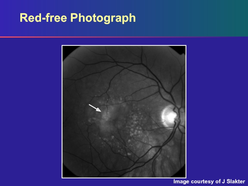
- 2. Red-free PhotographImage courtesy of J Slakter
- 3. Red-free PhotographRed-free photograph: Shallow subretinal fluidDrusenNote the
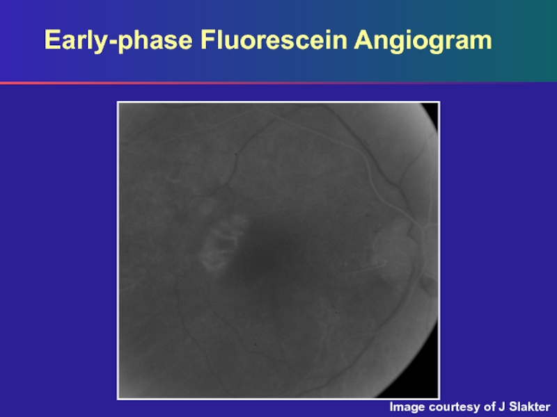
- 4. Early-phase Fluorescein AngiogramImage courtesy of J Slakter
- 5. Early-phase Fluorescein AngiogramEarly (arterial) phase:The most important
- 6. Mid-phase Fluorescein AngiogramImage courtesy of J Slakter
- 7. Mid-phase Fluorescein AngiogramMid-phase:Well-circumscribed hyperfluorescent area with curved
- 8. Mid-phase Fluorescein AngiogramBy only examining this image
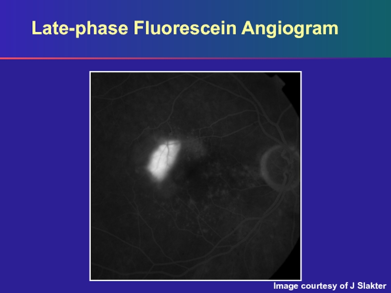
- 9. Late-phase Fluorescein AngiogramImage courtesy of J Slakter
- 10. Late-phase Fluorescein AngiogramLate phase:Increased hyperfluorescence with mild
- 11. Скачать презентанцию
Red-free PhotographImage courtesy of J Slakter
Слайды и текст этой презентации
Слайд 3Red-free Photograph
Red-free photograph:
Shallow subretinal fluid
Drusen
Note the area just temporal
Слайд 5Early-phase Fluorescein Angiogram
Early (arterial) phase:
The most important frame
Note the
oval area of mild hyperfluorescence occurring when the retinal arteries
are just fillingThe choroid is also just beginning to fill
Horizontally oriented vessels represent larger choroidal vessels seen through a rip of the RPE
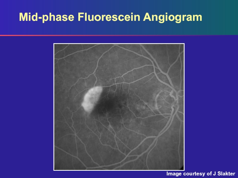
Слайд 7Mid-phase Fluorescein Angiogram
Mid-phase:
Well-circumscribed hyperfluorescent area with curved outer border and
almost straight line margin adjacent to an area of relative
hypofluorescenceFindings are typical of RPE rips and should act as clues to the diagnosis
Слайд 8Mid-phase Fluorescein Angiogram
By only examining this image and the one
that follows, this can be mistaken for classic CNV
The very
early image helps to make the correct diagnosisThis can also be misdiagnosed as geographic atrophy, which has:
A border that is geographic not linear
A lesion with no overlying retinal detachment

