Разделы презентаций
- Разное
- Английский язык
- Астрономия
- Алгебра
- Биология
- География
- Геометрия
- Детские презентации
- Информатика
- История
- Литература
- Математика
- Медицина
- Менеджмент
- Музыка
- МХК
- Немецкий язык
- ОБЖ
- Обществознание
- Окружающий мир
- Педагогика
- Русский язык
- Технология
- Физика
- Философия
- Химия
- Шаблоны, картинки для презентаций
- Экология
- Экономика
- Юриспруденция
SIW Theme : “Benign tumor of the external genitalia”
Содержание
- 1. SIW Theme : “Benign tumor of the external genitalia”
- 2. PlanIntroductionEtiology and pathogenesisClassificationConclusionReferences
- 3. IntroductionBenign tumors are characterized by slow growth,
- 4. The types of benign tumors of external genital organs:Fibroma(fibroids)LipomaMyxomaMyomaHemangiomaLymphangiomaPapillomaHidradenoma
- 5. Etiology and pathogenesis The process of tumor
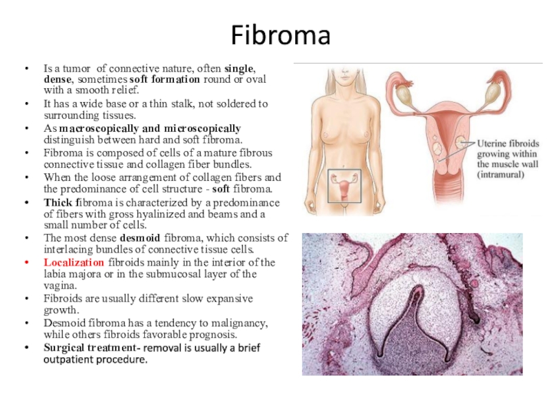
- 6. FibromaIs a tumor of connective nature, often
- 7. Fibroidsswelling of the muscle fibers, often comes
- 8. Lipomadevelops mainly of fat tissue in the
- 9. Myxomatumor that develops from the mesenchyme residues
- 10. MyomaTumor from muscle fibers, often comes from
- 11. Pathogenesis of myomain the course of repeatedly
- 12. ClinicsUterine bleeding as metro- or menorrhagia with
- 13. DiagnosticsAnamnesis collection Bimanual examination Ultrasound examination using
- 14. TreatmentMedicationSurgery (abdominal, laparoscopic myomectomy) B. radical surgery
- 15. MedicationAnalogues of the releasing hormoneAntiprogestagens (mifepristone)AntigonadotropinsProgestogens (when combined with hyperplastic endometrial processes) COC Non-hormonal symptomatic treatment
- 16. Hemangiomaoccurs on the basis of a congenital
- 17. Etiological factorsGirls have hemangiomas 2-3 times more
- 18. DiagnosticsExaminationLaboratory diagnosticsUltrasound examination (determining the depth of
- 19. TreatmentSurgical treatment is indicated for deep-seated vascular
- 20. Lymphangiomadevelops from the lymphatic vessels of the
- 21. Papillomaswelling of the surface epithelium, having the
- 22. Hidradenomaa benign tumor originating from the elements
- 23. ConclusionAnd in conclusion I want to say
- 24. ReferencesZh.Abissatov “Clinical oncogynecology” 2007y.( p.618-626)http://upladdy.com/en/pages/1602940https://myslide.ru/presentation/skachat-dobrokachestvennye-novoobrazovaniya-zhenskix-polovyx-organov
- 25. Скачать презентанцию
PlanIntroductionEtiology and pathogenesisClassificationConclusionReferences
Слайды и текст этой презентации
Слайд 4The types of benign tumors of external genital organs:
Fibroma(fibroids)
Lipoma
Myxoma
Myoma
Hemangioma
Lymphangioma
Papilloma
Hidradenoma
Слайд 5Etiology and pathogenesis
The process of tumor transformation of cells
has not yet been fully studied. It is based on
damage to the genetic material of the cell (DNA), leading to disruption in the mechanisms of cell division and growth control, as well as mechanisms of apoptosis (programmed cell death). At the moment, there are a lot of factors that can cause such changes in normal cells:Chemical factors: polycyclic aromatic hydrocarbons and other chemical substances of an aromatic nature are able to react with the DNA of cells, damaging it.
Physical factors: ultraviolet radiation and other types of ionizing radiation damage cellular structures (including DNA), causing tumor transformation of cells.
Mechanical injuries and high temperatures with long-term exposure to the body contribute to the process of carcinogenesis.
Biological factors are mainly viruses. At the moment, the leading role of the human papilloma virus in the development of cervical cancer has been proven.
Disturbance of immune system function is the main cause of tumor development in patients with a decreased function of the immune system (AIDS patients).
Violation of the endocrine system. A large number of tumors develops as a result of a violation of the body's hormonal balance
Слайд 6Fibroma
Is a tumor of connective nature, often single, dense, sometimes
soft formation round or oval with a smooth relief.
It has
a wide base or a thin stalk, not soldered to surrounding tissues.As macroscopically and microscopically distinguish between hard and soft fibroma.
Fibroma is composed of cells of a mature fibrous connective tissue and collagen fiber bundles.
When the loose arrangement of collagen fibers and the predominance of cell structure - soft fibroma.
Thick fibroma is characterized by a predominance of fibers with gross hyalinized and beams and a small number of cells.
The most dense desmoid fibroma, which consists of interlacing bundles of connective tissue cells.
Localization fibroids mainly in the interior of the labia majora or in the submucosal layer of the vagina.
Fibroids are usually different slow expansive growth.
Desmoid fibroma has a tendency to malignancy, while others fibroids favorable prognosis.
Surgical treatment- removal is usually a brief outpatient procedure.
Слайд 7Fibroids
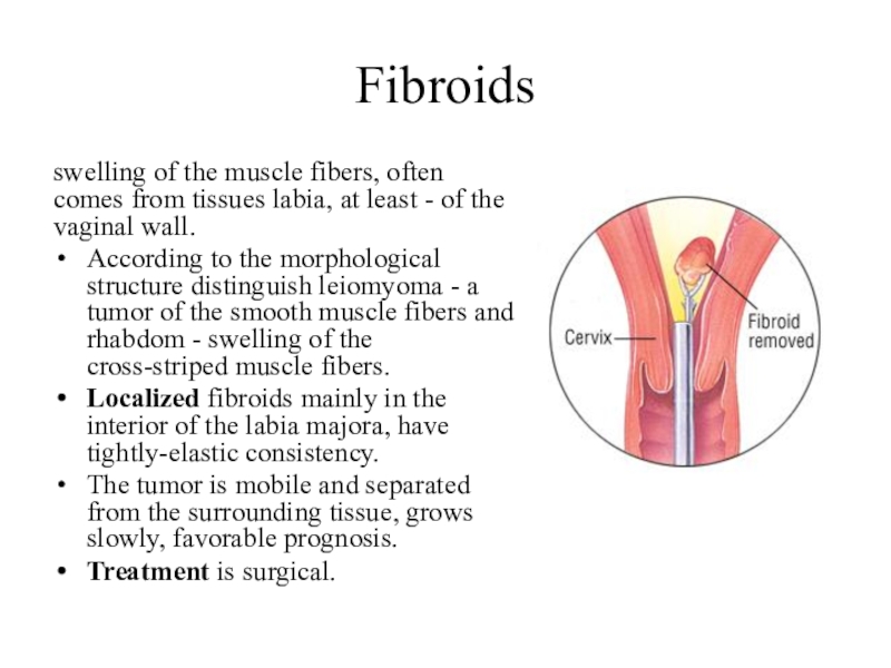
swelling of the muscle fibers, often comes from tissues labia,
at least - of the vaginal wall.
According to the morphological
structure distinguish leiomyoma - a tumor of the smooth muscle fibers and rhabdom - swelling of the cross-striped muscle fibers.Localized fibroids mainly in the interior of the labia majora, have tightly-elastic consistency.
The tumor is mobile and separated from the surrounding tissue, grows slowly, favorable prognosis.
Treatment is surgical.
Слайд 8Lipoma
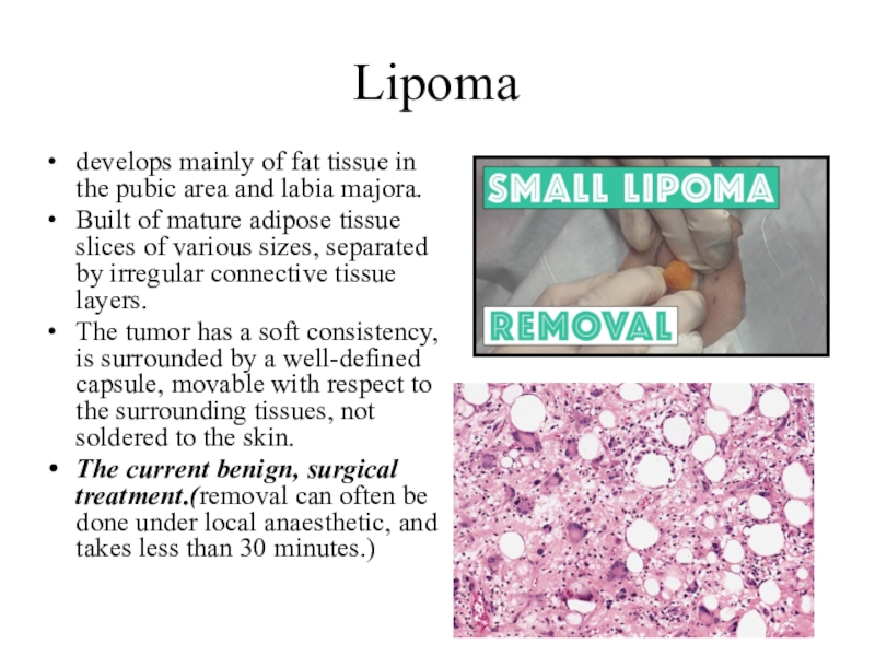
develops mainly of fat tissue in the pubic area and
labia majora.
Built of mature adipose tissue slices of various sizes,
separated by irregular connective tissue layers.The tumor has a soft consistency, is surrounded by a well-defined capsule, movable with respect to the surrounding tissues, not soldered to the skin.
The current benign, surgical treatment.(removal can often be done under local anaesthetic, and takes less than 30 minutes.)
Слайд 9Myxoma
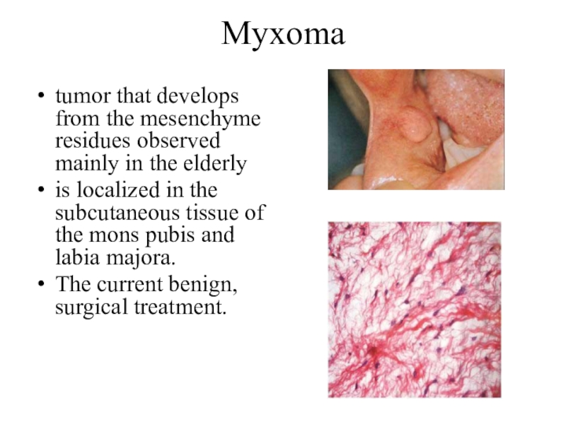
tumor that develops from the mesenchyme residues observed mainly in
the elderly
is localized in the subcutaneous tissue of the mons
pubis and labia majora.The current benign, surgical treatment.
Слайд 10Myoma
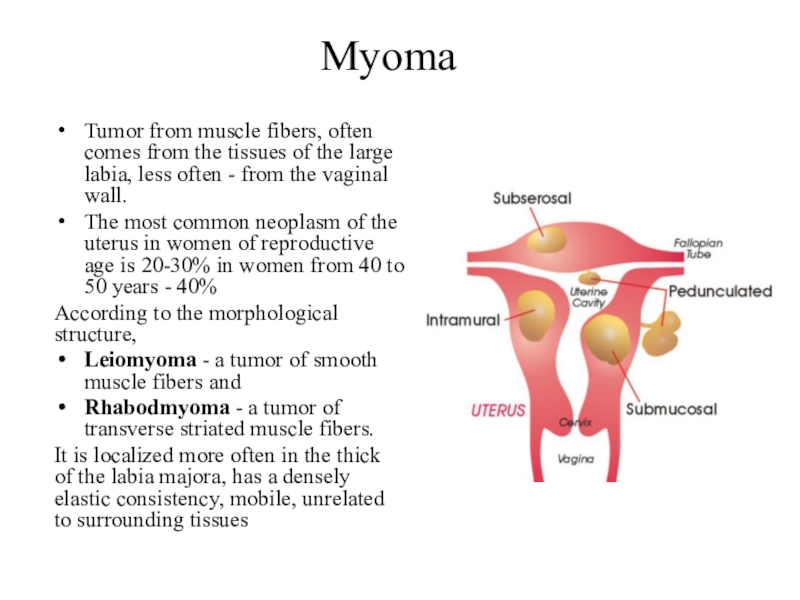
Tumor from muscle fibers, often comes from the tissues of
the large labia, less often - from the vaginal wall.
The
most common neoplasm of the uterus in women of reproductive age is 20-30% in women from 40 to 50 years - 40%According to the morphological structure,
Leiomyoma - a tumor of smooth muscle fibers and
Rhabodmyoma - a tumor of transverse striated muscle fibers.
It is localized more often in the thick of the labia majora, has a densely elastic consistency, mobile, unrelated to surrounding tissues
Слайд 11Pathogenesis of myoma
in the course of repeatedly repeated cycles of
myometrium hyperplasia followed by apoptosis
(the dynamics of the normal menstrual cycle) there is an accumulation of smooth muscle cells with disturbed apoptosis during, these proliferating cells are exposed to various damaging factors:- ischemia (including during menstruation)
- inflamed a process - a traumatic effect due to medical manipulations
- the focus of endometriosis
Слайд 12Clinics
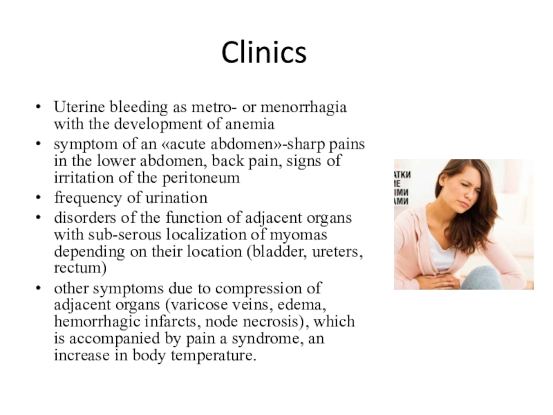
Uterine bleeding as metro- or menorrhagia with the development of
anemia
symptom of an «acute abdomen»-sharp pains in the lower abdomen,
back pain, signs of irritation of the peritoneumfrequency of urination
disorders of the function of adjacent organs with sub-serous localization of myomas depending on their location (bladder, ureters, rectum)
other symptoms due to compression of adjacent organs (varicose veins, edema, hemorrhagic infarcts, node necrosis), which is accompanied by pain a syndrome, an increase in body temperature.
Слайд 13Diagnostics
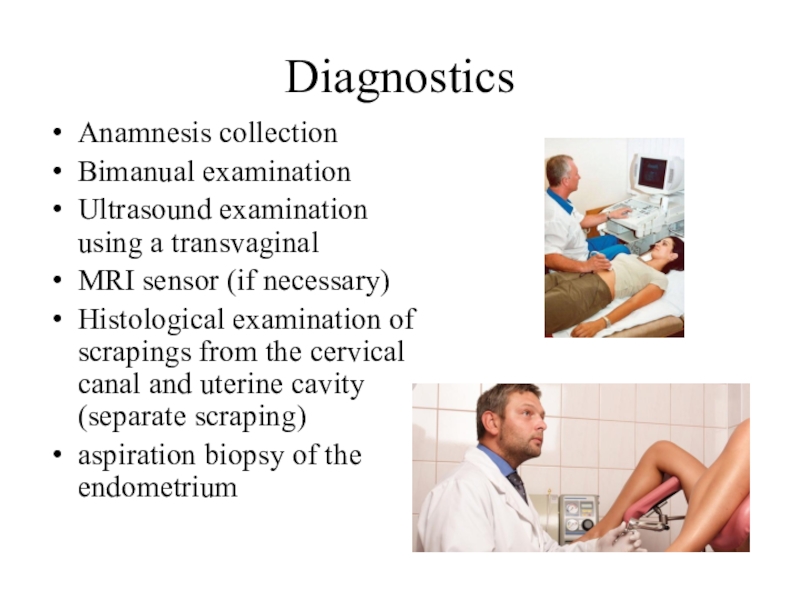
Anamnesis collection
Bimanual examination
Ultrasound examination using a transvaginal
MRI
sensor (if necessary)
Histological examination of scrapings from the cervical canal
and uterine cavity (separate scraping)aspiration biopsy of the endometrium
Слайд 14Treatment
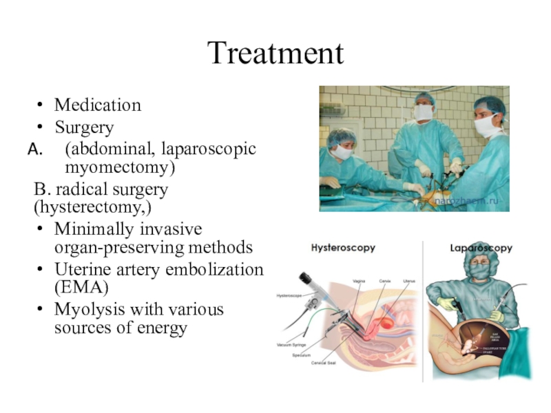
Medication
Surgery
(abdominal, laparoscopic myomectomy)
B. radical surgery (hysterectomy,)
Minimally invasive
organ-preserving methods
Uterine artery embolization (EMA)
Myolysis with various sources of
energyСлайд 15Medication
Analogues of the releasing hormone
Antiprogestagens (mifepristone)
Antigonadotropins
Progestogens (when combined with hyperplastic
endometrial processes)
COC
Non-hormonal symptomatic treatment
Слайд 16Hemangioma
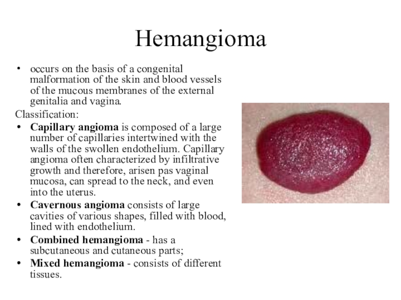
occurs on the basis of a congenital malformation of the
skin and blood vessels of the mucous membranes of the
external genitalia and vagina.Classification:
Capillary angioma is composed of a large number of capillaries intertwined with the walls of the swollen endothelium. Capillary angioma often characterized by infiltrative growth and therefore, arisen pas vaginal mucosa, can spread to the neck, and even into the uterus.
Cavernous angioma consists of large cavities of various shapes, filled with blood, lined with endothelium.
Combined hemangioma - has a subcutaneous and cutaneous parts;
Mixed hemangioma - consists of different tissues.
Слайд 17Etiological factors
Girls have hemangiomas 2-3 times more often than boys.
Usually
they are detected immediately after birth and much less often
- during the first two months of life.The preferred location of hemangiomas is the area of the head and neck. The most rare localization in the head and neck region is the parotid region.
Hemangiomas can be single and multiple.
A feature of hemangiomas in the perineum and external genitalia is their tendency to frequent ulceration, which leads to self-healing of the tumor.
Hemangiomas is defined as bluish, purple, and red spots, a few rise above the level of the mucous membrane or skin.
The mucous membrane in the swollen spots, wrinkles her raw, surface epithelium loosened.
Colposcopic determined by an extensive network of anastomosing, sometimes varicose vessels.
The tumor may reach large sizes and distributed on the internal genitalia.
Слайд 18Diagnostics
Examination
Laboratory diagnostics
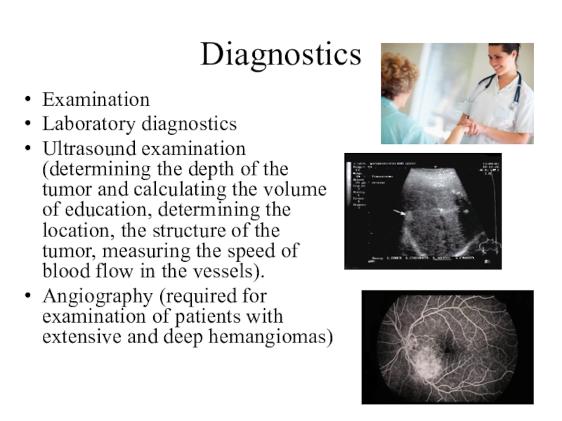
Ultrasound examination (determining the depth of the tumor and
calculating the volume of education, determining the location, the structure
of the tumor, measuring the speed of blood flow in the vessels).Angiography (required for examination of patients with extensive and deep hemangiomas)
Слайд 19Treatment
Surgical treatment is indicated for deep-seated vascular hemangiomas, when it
is possible to remove the entire formation, within the limits
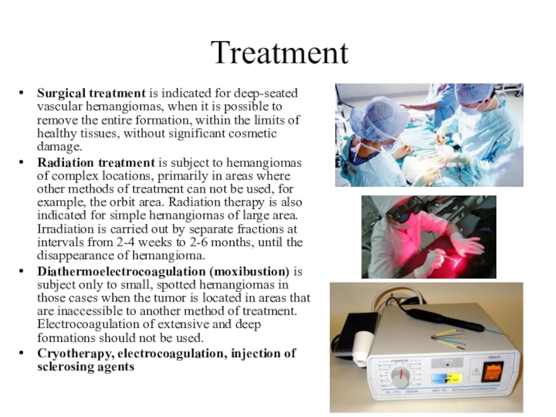
of healthy tissues, without significant cosmetic damage.Radiation treatment is subject to hemangiomas of complex locations, primarily in areas where other methods of treatment can not be used, for example, the orbit area. Radiation therapy is also indicated for simple hemangiomas of large area. Irradiation is carried out by separate fractions at intervals from 2-4 weeks to 2-6 months, until the disappearance of hemangioma.
Diathermoelectrocoagulation (moxibustion) is subject only to small, spotted hemangiomas in those cases when the tumor is located in areas that are inaccessible to another method of treatment. Electrocoagulation of extensive and deep formations should not be used.
Cryotherapy, electrocoagulation, injection of sclerosing agents
Слайд 20Lymphangioma
develops from the lymphatic vessels of the skin most often
in areas close to the groin.
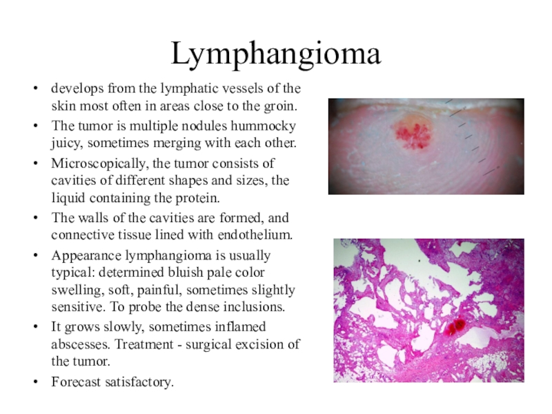
The tumor is multiple nodules
hummocky juicy, sometimes merging with each other.Microscopically, the tumor consists of cavities of different shapes and sizes, the liquid containing the protein.
The walls of the cavities are formed, and connective tissue lined with endothelium.
Appearance lymphangioma is usually typical: determined bluish pale color swelling, soft, painful, sometimes slightly sensitive. To probe the dense inclusions.
It grows slowly, sometimes inflamed abscesses. Treatment - surgical excision of the tumor.
Forecast satisfactory.
Слайд 21Papilloma
swelling of the surface epithelium, having the form of papillary
proliferation, with exophytic growth often in the labia majora, at
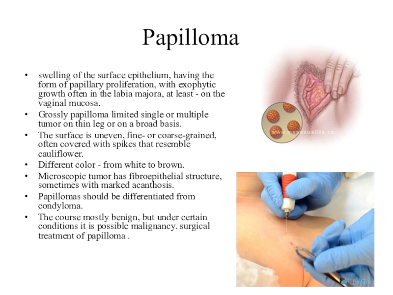
least - on the vaginal mucosa.Grossly papilloma limited single or multiple tumor on thin leg or on a broad basis.
The surface is uneven, fine- or coarse-grained, often covered with spikes that resemble cauliflower.
Different color - from white to brown.
Microscopic tumor has fibroepithelial structure, sometimes with marked acanthosis.
Papillomas should be differentiated from condyloma.
The course mostly benign, but under certain conditions it is possible malignancy. surgical treatment of papilloma .
Слайд 22Hidradenoma
a benign tumor originating from the elements of the sweat
glands.
Manifested mainly aged 15 to 20 years.
Localization - large labia
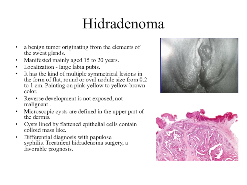
pubis.It has the kind of multiple symmetrical lesions in the form of flat, round or oval nodule size from 0.2 to 1 cm. Painting on pink-yellow to yellow-brown color.
Reverse development is not exposed, not malignant .
Microscopic cysts are defined in the upper part of the dermis.
Cysts lined by flattened epithelial cells contain colloid mass like.
Differential diagnosis with papulose syphilis. Treatment hidradenoma surgery, a favorable prognosis.


