Слайд 2Normal Pregnancy
Pregnancy
The course that the embryo and
the fetus grow in the maternal body
Stages of pregnancy
Early pregnancy:
≤12 weeks
Mid pregnancy: ≥13 weeks,≤27 weeks
Late pregnancy:≥28 weeks
Term pregnancy:≥37 weeks,<42 weeks
Слайд 3Formation of Embryo
Fertilization
Place: oviduct (ampulla)
Process
capacitation →
acrosome reaction→ penetrate the zona pellucida→ second meiosis →zygote
Слайд 4Formation of Embryo
Implantation
requirement
Disappear of zona pellucida
Formation of syncytiotrophoblast
Synchronized development of
blastocyst and endometrium
Adequate progesterone
Слайд 5Formation of Embryo
Process
morula (day 3) → enter uterine cavity (day
4) → early blastocyst→ late blastocyst (day 6-7) → implantation
location→
adherence→ penetration
Слайд 6Development of embryo and fetus
Definition
embryo: ≤ 8 weeks
Fetus: ≥ 9
weeks, human shape
Слайд 7Development of embryo and fetus
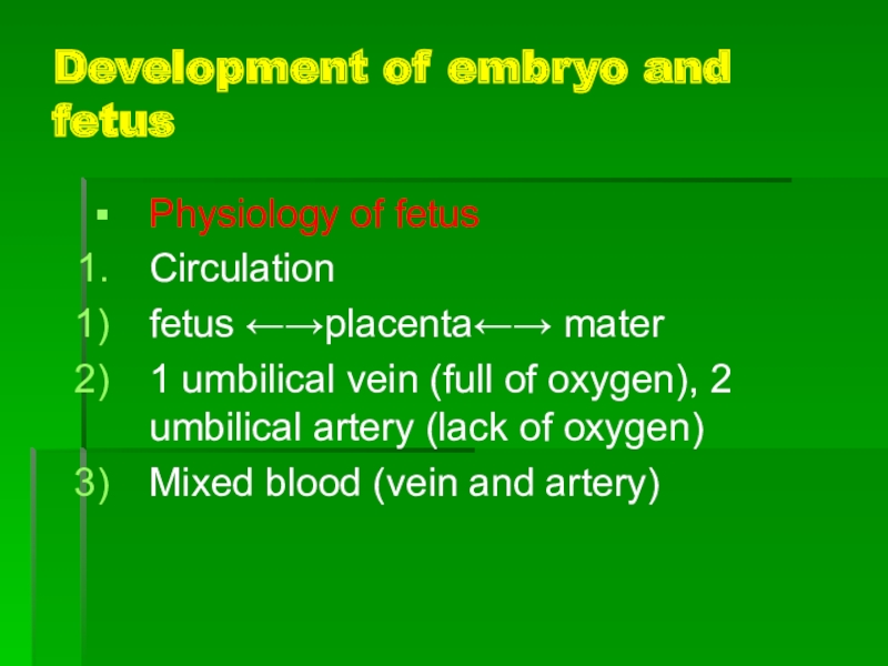
Physiology of fetus
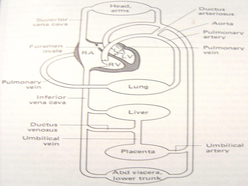
Circulation
fetus ←→placenta←→ mater
1 umbilical
vein (full of oxygen), 2 umbilical artery (lack of oxygen)
Mixed
blood (vein and artery)
Слайд 8Development of embryo and fetus
Слайд 9Development of embryo and fetus
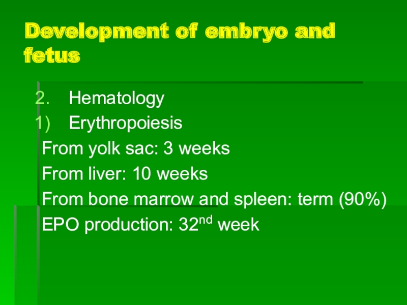
Hematology
Erythropoiesis
From yolk sac: 3 weeks
From liver:
10 weeks
From bone marrow and spleen: term (90%)
EPO production: 32nd
week
Слайд 10Development of embryo and fetus
Fetal hemoglobin
Fetal hemoglobin: early pregnancy
Adult hemoglobin:
32nd week
Term: fetal type Hb 25%
White cells
Leukocytes: 8 week
Lymphocytes (antibody
production): 12 week, thymus and spleen
Слайд 11Development of embryo and fetus
Gastrointestinal tract
drink amniotic fluid: 4th month
no
proteolytic activity
enzymatic deficiencies in liver:
bilirubin is not easy to
be clear.
Слайд 12Development of embryo and fetus
Kidney
Its function begins
at 11-14th week
Endocrinology
Fetal thyroid: the first endocrine gland (6th week),
synthesize thyroxine at 12th week
Fetal adrenal cortex: widen (20th week), a fetal zone. synthesize steroid hormones (E3, liver placenta mater)
Слайд 13Placenta
Structure
Primary villus
syncytiotrophoblast cytotrophoblast
Secondary villus
third class vilus
fetal capillary
enter the stroma
Слайд 14Placenta: Villi
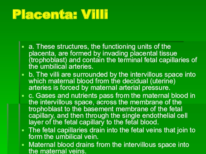
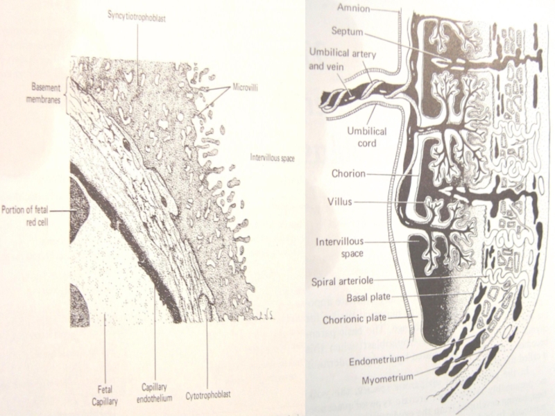
a. These structures, the functioning units of the
placenta, are formed by invading placental tissue (trophoblast) and contain
the terminal fetal capillaries of the umbilical arteries.
b. The villi are surrounded by the intervillous space into which maternal blood from the decidual (uterine) arteries is forced by maternal arterial pressure.
c. Gases and nutrients pass from the maternal blood in the intervillous space, across the membrane of the trophoblast to the basement membrane of the fetal capillary, and then through the single endothelial cell layer of the fetal capillary to the fetal blood.
The fetal capillaries drain into the fetal veins that join to form the umbilical vein.
Maternal blood drains from the intervillous space into the maternal veins.
Слайд 15Placenta: cotyledons
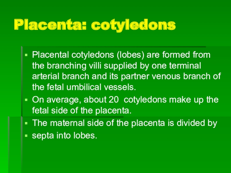
Placental cotyledons (lobes) are formed from the branching villi
supplied by one terminal arterial branch and its partner venous
branch of the fetal umbilical vessels.
On average, about 20 cotyledons make up the fetal side of the placenta.
The maternal side of the placenta is divided by
septa into lobes.
Слайд 17Placenta: structure
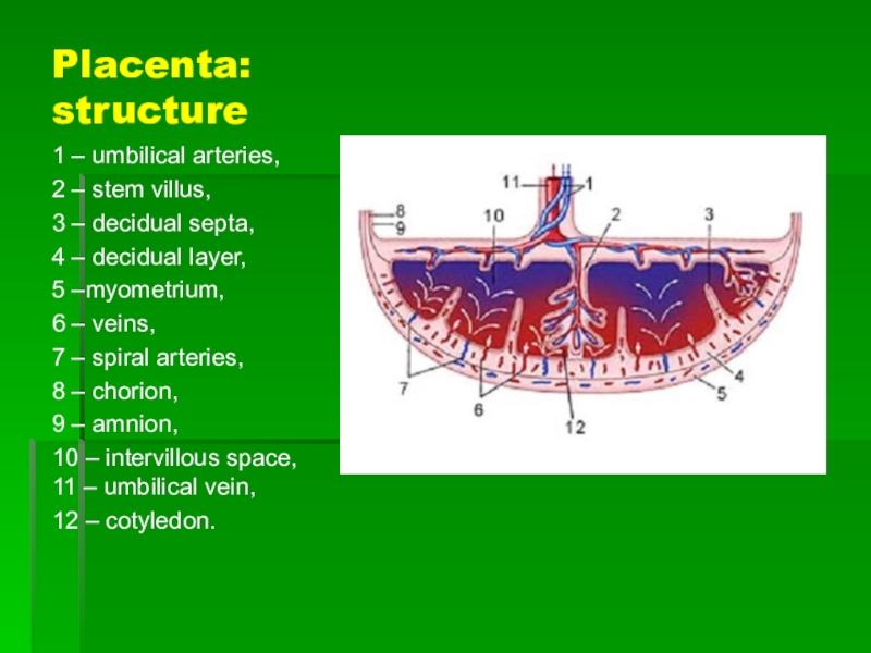
1 – umbilical arteries,
2 – stem villus,
3
– decidual septa,
4 – decidual layer,
5 –myometrium,
6
– veins,
7 – spiral arteries,
8 – chorion,
9 – amnion,
10 – intervillous space, 11 – umbilical vein,
12 – cotyledon.
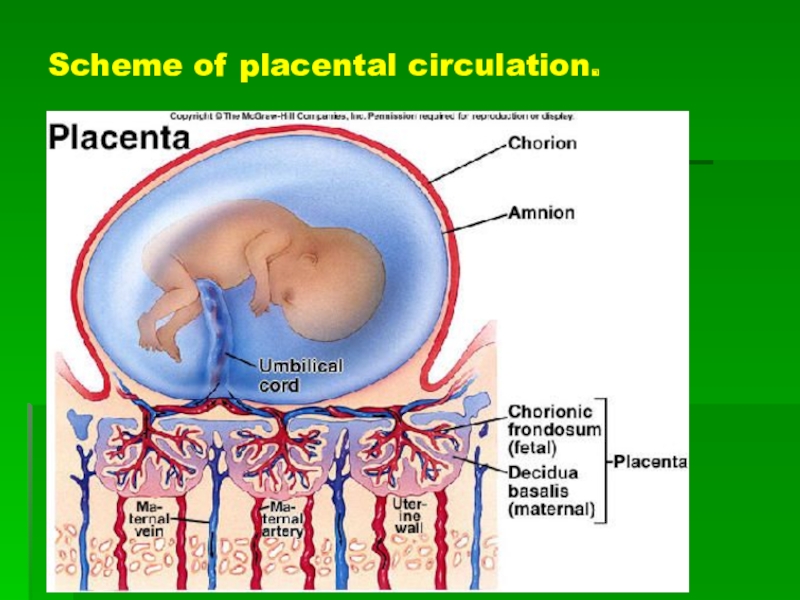
Слайд 18Scheme of placental circulation.
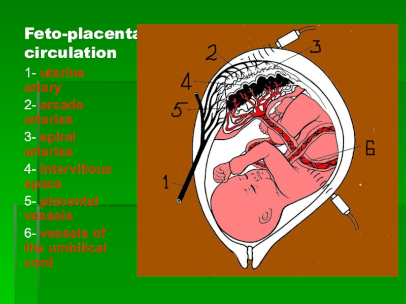
Слайд 20Feto-placental circulation
1- uterine artery
2- arcade arteries
3- spiral arteries
4- intervillous
space
5- placental vessels
6- vessels of the umbilical cord
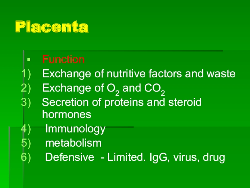
Слайд 21Placenta
Function
Exchange of nutritive factors and waste
Exchange of O2 and CO2
Secretion
of proteins and steroid hormones
Immunology
metabolism
Defensive - Limited.
IgG, virus, drug
Слайд 22Placenta: functions
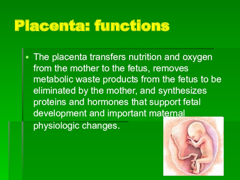
The placenta transfers nutrition and oxygen from the mother
to the fetus, removes metabolic waste products from the fetus
to be eliminated by the mother, and synthesizes proteins and hormones that support fetal development and important maternal physiologic changes.
Слайд 231. Mother-to-fetus transfer of nutrients
a. The essential substances for growth
and development move from the mother to the fetus in
four ways:
(1) Active transport: amino acids, calcium
(2) Facilitated transport: glucose
(3) Endocytosis: cholesterol, insulin, iron, immunoglobulin G (IgG)
(4) Sodium pumps and chloride channels: ions
b. Solute size and lipid solubility are also important factors that influence transport.
Слайд 242. Gas exchange
This process involves supplying oxygen to the fetus
and removing carbon dioxide
from the fetus.
Слайд 253. Secretion of proteins and steroid hormones
a. Progesterone is produced
by the placenta from maternal cholesterol, is secreted into the
maternal circulation, and is important for maintaining pregnancy.
b. Estrogen is converted from circulating fetal androgens (dehydroepiandrosterone sulfate [DHEAS] produced in the fetal adrenal glands. Estrogen plays an important role in maternal physiologic changes in pregnancy, labor, and lactation.
c. Numerous proteins, peptides, and growth factors are produced in the placenta. They are important for placental growth, fetal growth and development, and the maternal physiologic changes necessary to ensure adequate nutrition to the fetus.
Слайд 264. Immunology.
Invading placental cells express a unique antigen, HLA-G, which
is not recognized as a "foreign" antigen by the mother.
Other unique antigens and local immune suppression contribute to the prevention of rejection of the fetal-placental unit.
5. Metabolism. Glucose is the primary substrate for placental aerobic metabolism.
Слайд 27Fetal membranes
Structure
chorion and amnion
Amnion
A double-layered translucent
membrane
Become distended with fluid
Слайд 29Umbilical cord
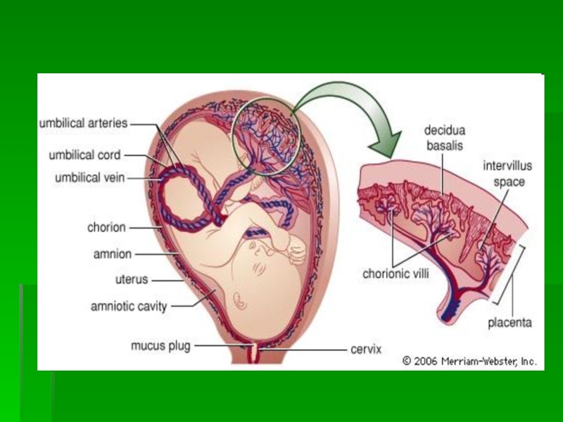
A. Umbilical arteries. Two umbilical arteries originate from the
fetal aorta. They supply fetal blood to all portions of
the placenta for gas and solute exchange. A single umbilical artery is associated with low birth weight and chromosomal anomalies in about 10 to 15% of infants.
B. Umbilical vein. One umbilical vein returns nutrient-rich, oxygen-rich blood to the fetus.
Wharton jelly
Umbilical cord Length - 30-70cm
Слайд 30Umbilical cord
In most cases, the cord is about 20 inches
long and almost 1 inch in diameter. It usually appears
loosely coiled. Inside the cord are two arteries and one vein. The vein supplies the baby with oxygenated, nutrient-rich blood, and the arteries carry de-oxygenated, nutrient-depleted blood back to the placenta. On occasion, the umbilical cord will only have two vessels; one artery and one vein.
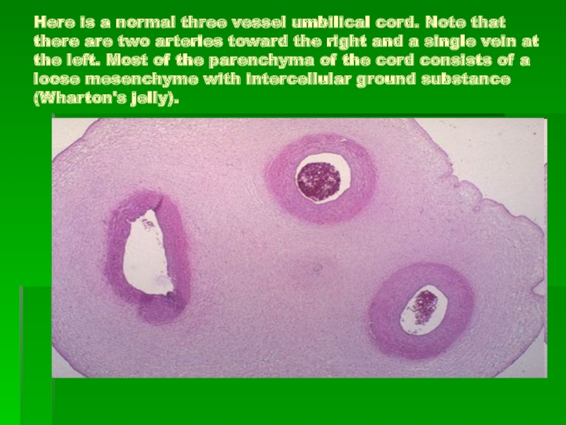
Слайд 31Here is a normal three vessel umbilical cord. Note that
there are two arteries toward the right and a single
vein at the left. Most of the parenchyma of the cord consists of a loose mesenchyme with intercellular ground substance (Wharton's jelly).
Слайд 33Amniotic fluid
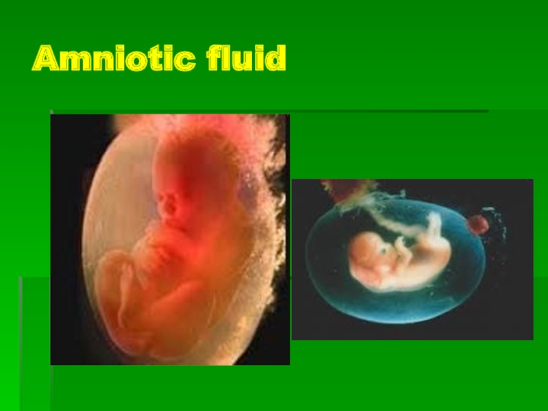
Amniotic fluid ( AF ) - the habitat of
the fetus, performs several functions simultaneously : the creation of
spaces for free movement of the growing fetus , protection from mechanical injury , maintaining temperature balance , preventing compression of the umbilical cord at birth , the implementation of the transport function and participation in metabolism .
AF is yellowish in early pregnancy, then lighter and transparent, and - cloudy , opalescent at the end of pregnancy ; pH - 6,98-7,23, specific gravity- 1007-1080 g / l , the protein content - 0.18-0.2 % glucose - 22 mg% urea - 23 mg%. AF may contain embryonic hair (lanugo), cells of the epidermis , sebaceous gland cells (vernix caseosa).
Слайд 34Amniotic fluid
AF volume depends on the term of pregnancy. Increase
in volume is uneven. The peak of AF volume fixed
at 33.8 weeks and is 931 ml. AF volume in the range 22-39 weeks does not change significantly (630 ml and 817 ml, respectively) and averaged 777 ml .
Слайд 35Amniotic Fliud
Towards the end pregnancy (term of labor) the volume
of amniotic fluid comes up to 1-1.5 liters, and every
three hours it is completely updated, with one-third recycled by fetus.
Слайд 36Amniotic Fluid Index (AFI)
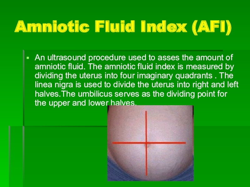
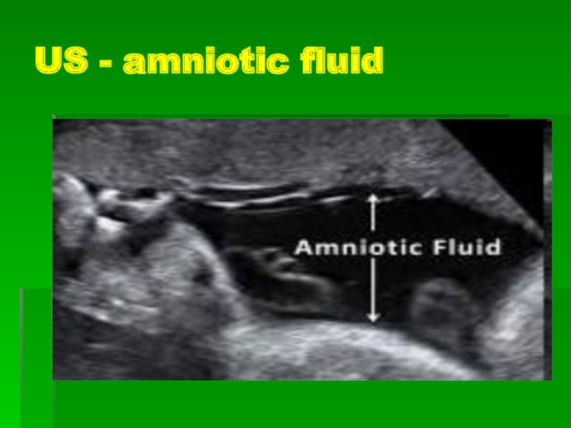
An ultrasound procedure used to asses the
amount of amniotic fluid. The amniotic fluid index is measured
by dividing the uterus into four imaginary quadrants . The linea nigra is used to divide the uterus into right and left halves.The umbilicus serves as the dividing point for the upper and lower halves.
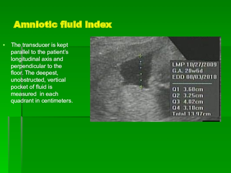
Слайд 37Amniotic fluid index
The transducer is kept parallel to the patient’s
longitudinal axis and perpendicular to the floor. The deepest, unobstructed,
vertical pocket of fluid is measured in each quadrant in centimeters.
Слайд 38AFI at different terms of pregnancy
(Amniotic Fluid Index Percentile Values)
Слайд 39Amniotic Fluid Index Percentile Values (mm)
Wks
2.5th 5th 50th 95th 97th
Слайд 41Amniotic Fliud
Function
Protect fetal
move freely, warm
Protect mater
prevent infection
Слайд 42Amniotic fliud
Source
exudation of fetal membranes (early pregnancy)
Fetal urine
Fetal lung
Exudation of
amnion and fetal skin
Слайд 43Amniotic fliud
Absord
Fetal membrane
Umbilical cord
Fetal skin
Fetal drinking
Feature
1000-1500ml at 36th-38th week (peak),
transparent → slightly turbid
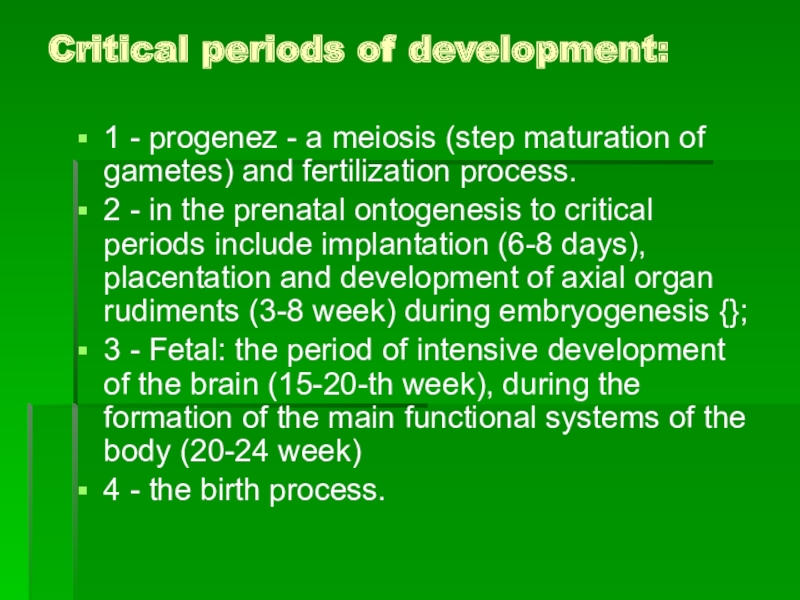
Слайд 44Critical periods of development:
1 - progenez - a meiosis
(step maturation of gametes) and fertilization process.
2 - in
the prenatal ontogenesis to critical periods include implantation (6-8 days), placentation and development of axial organ rudiments (3-8 week) during embryogenesis {};
3 - Fetal: the period of intensive development of the brain (15-20-th week), during the formation of the main functional systems of the body (20-24 week)
4 - the birth process.
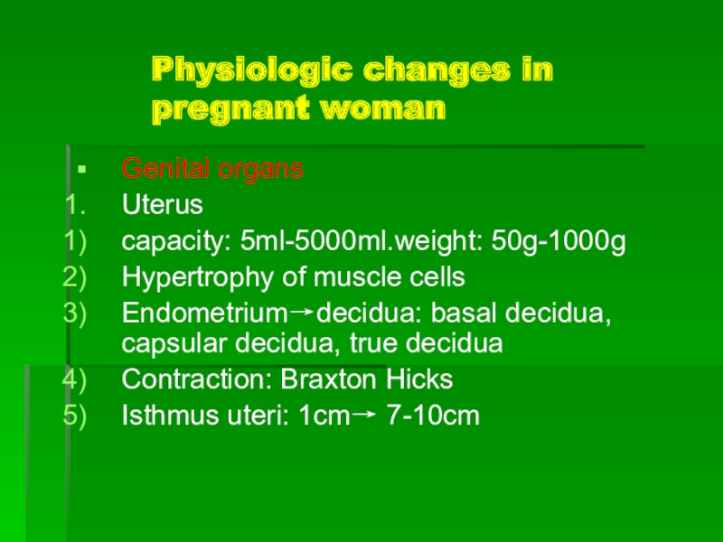
Слайд 45Physiologic changes in pregnant woman
Genital organs
Uterus
capacity: 5ml-5000ml.weight: 50g-1000g
Hypertrophy of muscle
cells
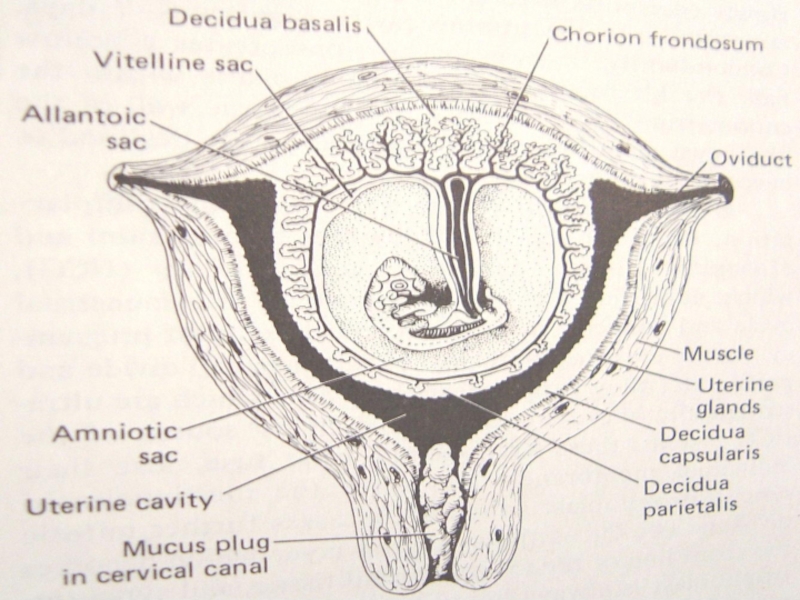
Endometrium→decidua: basal decidua, capsular decidua, true decidua
Contraction: Braxton Hicks
Isthmus uteri:
1cm→ 7-10cm
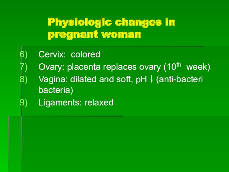
Слайд 47Physiologic changes in pregnant woman
Cervix: colored
Ovary: placenta replaces ovary (10th
week)
Vagina: dilated and soft, pH↓(anti-bacteri bacteria)
Ligaments: relaxed
Слайд 48Physiologic changes in pregnant woman
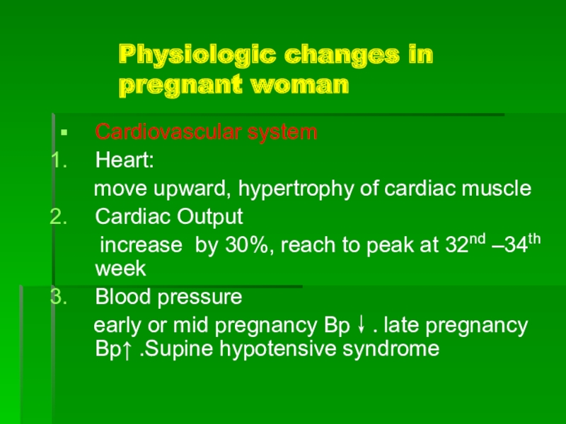
Cardiovascular system
Heart:
move upward,
hypertrophy of cardiac muscle
Cardiac Output
increase
by 30%, reach to peak at 32nd –34th week
Blood pressure
early or mid pregnancy Bp↓.late pregnancy Bp↑ .Supine hypotensive syndrome
Слайд 49Physiologic changes in pregnant woman
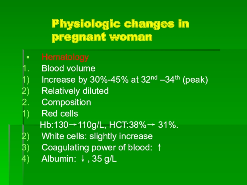
Hematology
Blood volume
Increase by 30%-45% at 32nd
–34th (peak)
Relatively diluted
Composition
Red cells
Hb:130→110g/L, HCT:38%→ 31%.
White cells:
slightly increase
Coagulating power of blood: ↑
Albumin: ↓,35 g/L
Слайд 50Physiologic changes in pregnant woman
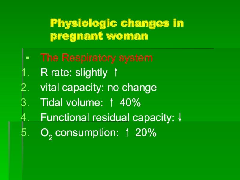
The Respiratory system
R rate: slightly ↑
vital
capacity: no change
Tidal volume: ↑ 40%
Functional residual capacity:↓
O2 consumption:
↑ 20%
Слайд 51Physiologic changes in pregnant woman
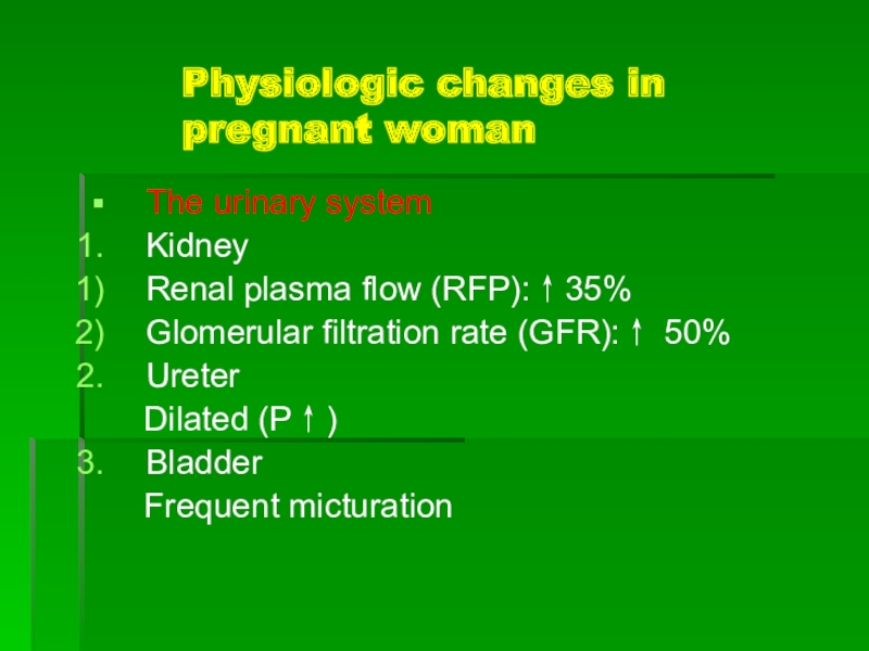
The urinary system
Kidney
Renal plasma flow (RFP):↑35%
Glomerular
filtration rate (GFR):↑ 50%
Ureter
Dilated (P↑)
Bladder
Frequent micturation
Слайд 52Physiologic changes in pregnant woman
Gastrointestinal system
Gastric emptying time is prolonged→
nausea.
The motility of large bowel is diminished → constipation
Liver
function: unchanged
Слайд 53Physiologic changes in pregnant woman
Endocrine
Pituitary (hypertrophy)
LH/FSH: ↓
PRL:↑
TSH and ACTH:↑
Thyroid
enlarged (TSH
and HCG↑)
thyroxine↑ and TBG↑ → free T3 T4 unchanged
Слайд 54Diagnosis of pregnancy
Questionable signs of pregnancy
Probable signs
True signs
Laboratory tests :
β-HCG, ptrogesterone
Additional methods : US
Слайд 55Questionable signs of pregnancy
Change of appetite.
Changes of smell (aversion
to perfume, tobacco, any other smells).
Changes of the nervous system:
quick fatigability, sleepiness, irritability, quick change of mood (instability of mood).
Слайд 56Questionable signs
Morning sickness.
Pigmentation of the skin ( nipple and
areolae, linea alba, forehead and cheeks).
Increase of fatty tissue,
enlargement of abdomen.
Frequency of micturition- due to: 1) pressure of the bulky uterus on the fundus of the bladder because of excessive anteverted position of the uterus; 2) congestion of the bladder mucous membrane, 3) stretching of the bladder base due to backward displacement of the cervix.
Breast discomfort.
Слайд 57Probable signs
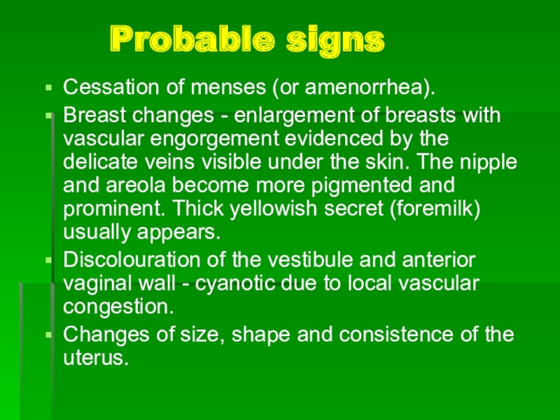
Cessation of menses (or amenorrhea).
Breast changes - enlargement
of breasts with vascular engorgement evidenced by the delicate veins
visible under the skin. The nipple and areola become more pigmented and prominent. Thick yellowish secret (foremilk) usually appears.
Discolouration of the vestibule and anterior vaginal wall - cyanotic due to local vascular congestion.
Changes of size, shape and consistence of the uterus.
Слайд 58Pregnancy’ sign in VE:
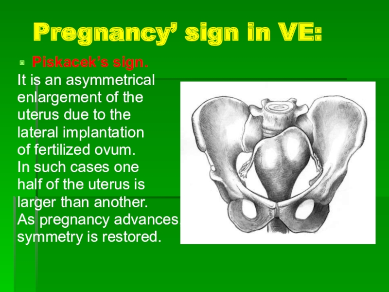
Piskacek’s sign.
It is an asymmetrical
enlargement
of the
uterus due to the
lateral implantation
of fertilized
ovum.
In such cases one
half of the uterus is
larger than another.
As pregnancy advances,
symmetry is restored.
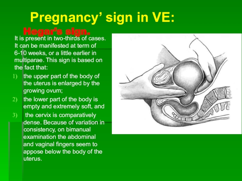
Слайд 59Hegar’s sign.
It is present in two-thirds of cases. It can
be manifested at term of 6-10 weeks, or a little
earlier in multiparae. This sign is based on the fact that:
the upper part of the body of the uterus is enlarged by the growing ovum;
the lower part of the body is empty and extremely soft, and
the cervix is comparatively dense. Because of variation in consistency, on bimanual examination the abdominal and vaginal fingers seem to appose below the body of the uterus.
Pregnancy’ sign in VE:
Слайд 60
Pregnancy’ sign in VE:
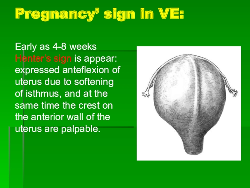
Early as 4-8 weeks Henter’s sign is
appear: expressed anteflexion of uterus due to softening of isthmus,
and at the same time the crest on the anterior wall of the uterus are palpable.
Слайд 61Pregnancy’ sign in VE:
Haus-Gubarev’s sign - the cervix of the
uterus becomes very mobile, due to softening of the isthmus
of the uterus.
Snegiryov’s sign – Increased irritability of the uterus body presented with appearance of hypertonicity of the uterus under palpating fingers during bimanual examination.
Слайд 62Uterus sizes
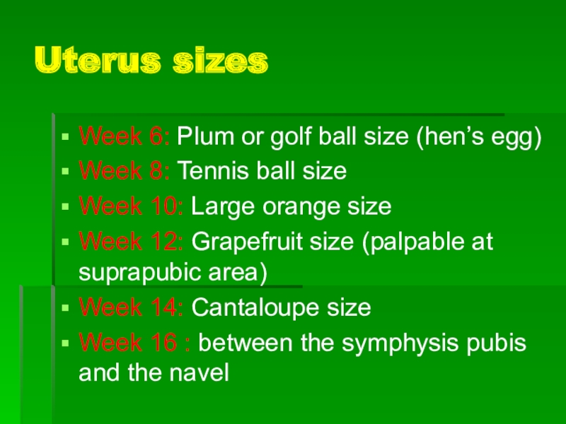
Week 6: Plum or golf ball size (hen’s egg)
Week
8: Tennis ball size
Week 10: Large orange size
Week 12: Grapefruit
size (palpable at suprapubic area)
Week 14: Cantaloupe size
Week 16 : between the symphysis pubis and the navel
Слайд 63Uterus sizes
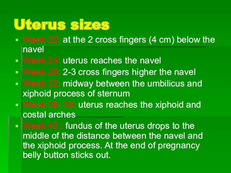
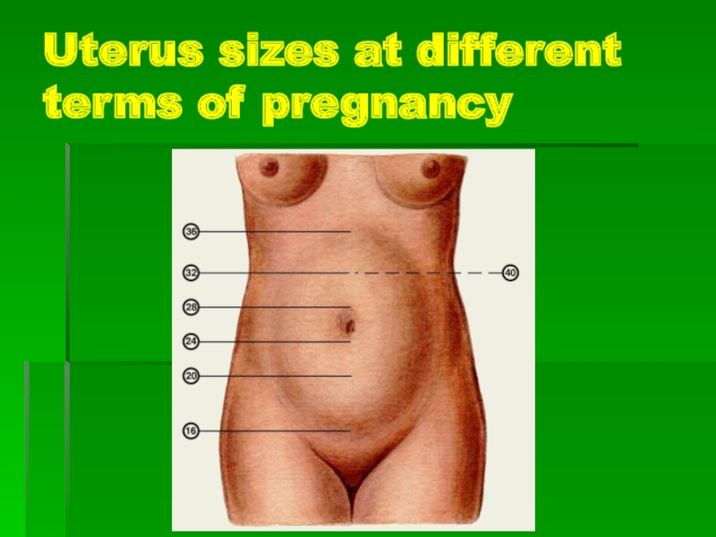
Week 20: at the 2 cross fingers (4 cm)
below the navel
Week 24: uterus reaches the navel
Week 28:
2-3 cross fingers higher the navel
Week 32: midway between the umbilicus and xiphoid process of sternum
Week 36- 38: uterus reaches the xiphoid and costal arches
Week 40 : fundus of the uterus drops to the middle of the distance between the navel and the xiphoid process. At the end of pregnancy belly button sticks out.
Слайд 64Uterus sizes at different terms of pregnancy
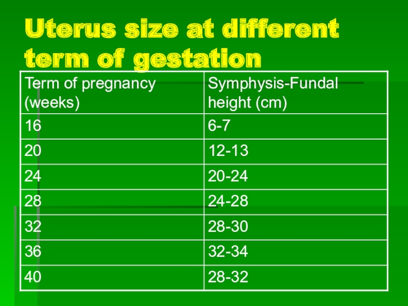
Слайд 65Uterus size at different term of gestation
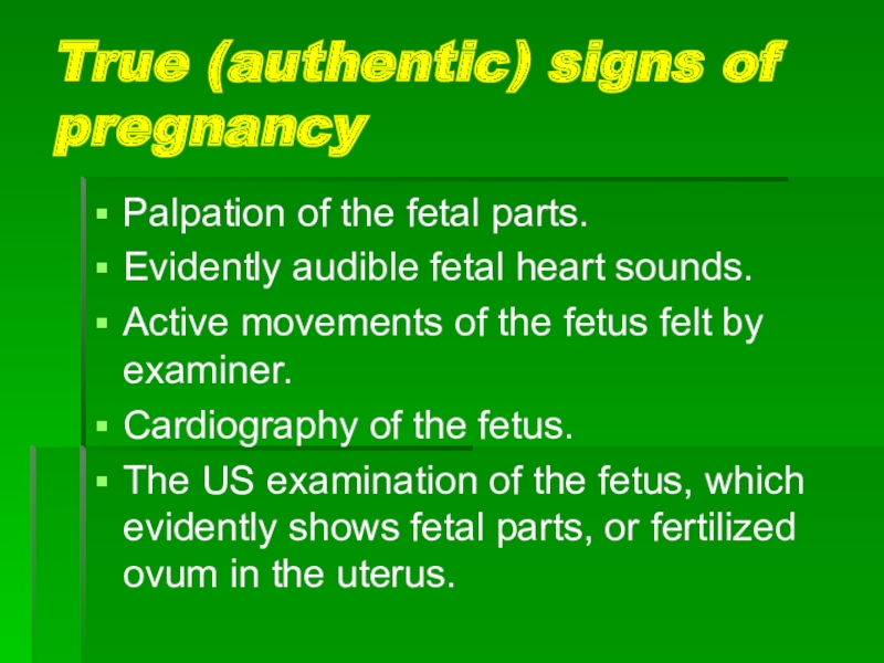
Слайд 66True (authentic) signs of pregnancy
Palpation of the fetal parts.
Evidently audible
fetal heart sounds.
Active movements of the fetus felt by examiner.
Cardiography
of the fetus.
The US examination of the fetus, which evidently shows fetal parts, or fertilized ovum in the uterus.
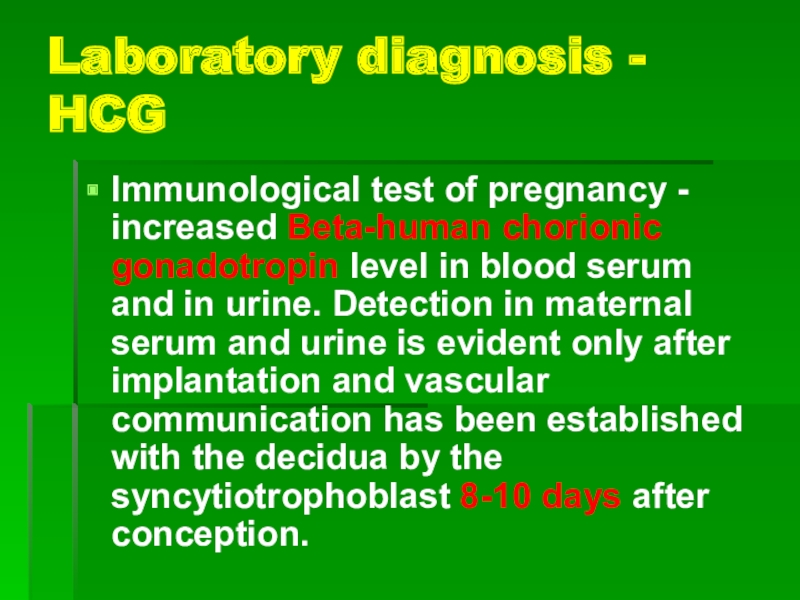
Слайд 67Laboratory diagnosis - HCG
Immunological test of pregnancy - increased Beta-human
chorionic gonadotropin level in blood serum and in urine. Detection
in maternal serum and urine is evident only after implantation and vascular communication has been established with the decidua by the syncytiotrophoblast 8-10 days after conception.
Слайд 68
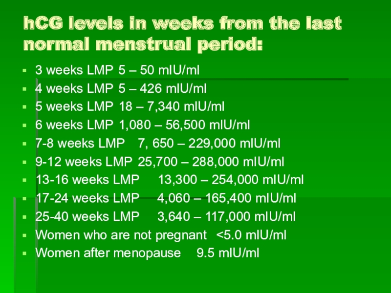
hCG levels in weeks from the last normal menstrual period:
3
weeks LMP 5 – 50 mIU/ml
4 weeks LMP 5 – 426 mIU/ml
5
weeks LMP 18 – 7,340 mIU/ml
6 weeks LMP 1,080 – 56,500 mIU/ml
7-8 weeks LMP 7, 650 – 229,000 mIU/ml
9-12 weeks LMP 25,700 – 288,000 mIU/ml
13-16 weeks LMP 13,300 – 254,000 mIU/ml
17-24 weeks LMP 4,060 – 165,400 mIU/ml
25-40 weeks LMP 3,640 – 117,000 mIU/ml
Women who are not pregnant <5.0 mIU/ml
Women after menopause 9.5 mIU/ml
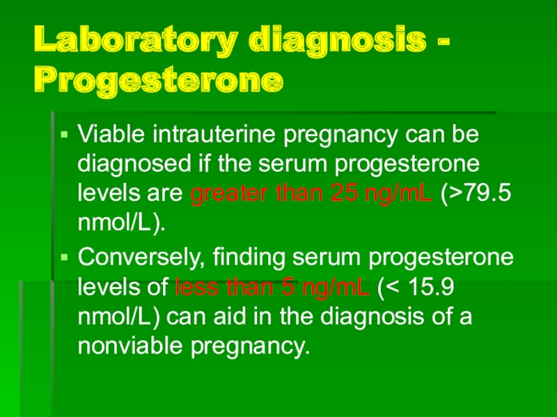
Слайд 69Laboratory diagnosis - Progesterone
Viable intrauterine pregnancy can be diagnosed if
the serum progesterone levels are greater than 25 ng/mL (>79.5
nmol/L).
Conversely, finding serum progesterone levels of less than 5 ng/mL (< 15.9 nmol/L) can aid in the diagnosis of a nonviable pregnancy.
Слайд 70Pregnancy diagnosis: Sonography
Transvaginal ultrasonography (TVUS), and transabdominal ultrasonography (TAUS) are
used to determine:
the fertiliezed ovum in the uterinbe cavity,
the size of the uterus (term of gestation),
cardiac motion can sometimes be identified in a 2- to 3-mm embryo but is almost always present when the embryo grows to 5 mm or longer. At 5-6 weeks' gestation, the fetal heart rate ranges from 100-115 beats per minute. At 9 week of gestation the heart rate ranges from 140 bpm.
Слайд 71Laboratory diagnosis - Progesterone
Viable intrauterine pregnancy can be diagnosed if
the serum progesterone levels are greater than 25 ng/mL (>79.5
nmol/L).
Conversely, finding serum progesterone levels of less than 5 ng/mL (< 15.9 nmol/L) can aid in the diagnosis of a nonviable pregnancy.
Слайд 72US exam
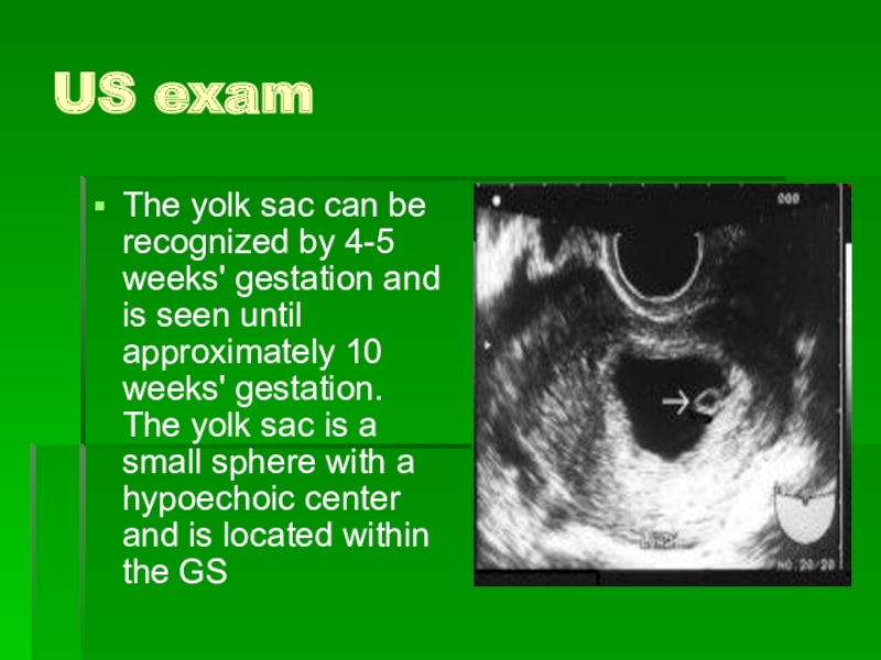
The yolk sac can be recognized by 4-5 weeks'
gestation and is seen until approximately 10 weeks' gestation. The
yolk sac is a small sphere with a hypoechoic center and is located within the GS

