Разделы презентаций
- Разное
- Английский язык
- Астрономия
- Алгебра
- Биология
- География
- Геометрия
- Детские презентации
- Информатика
- История
- Литература
- Математика
- Медицина
- Менеджмент
- Музыка
- МХК
- Немецкий язык
- ОБЖ
- Обществознание
- Окружающий мир
- Педагогика
- Русский язык
- Технология
- Физика
- Философия
- Химия
- Шаблоны, картинки для презентаций
- Экология
- Экономика
- Юриспруденция
Up-to-date diagnostic methods
Содержание
- 1. Up-to-date diagnostic methods
- 2. Up-to-date diagnostic methods
- 3. Optical Coherence TomographyStratus OCTNon-invasive optical diagnostic techniques
- 4. Optical Coherence Tomography of anterior eye segmentVisante OCT
- 5. OCT of anterior eye segmentprovides high-resolution anterior
- 6. The Non-Mydriatic Fundus Camera System from Carl Zeiss
- 7. The Non-Mydriatic Fundus CameraBrilliant true color retinal
- 8. Ultrasound eye examination
- 9. Fluorescein angiography
- 10. Ophthalmosurgery
- 11. Operating- room
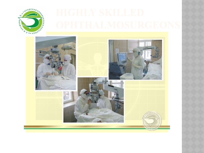
- 12. Highly skilled ophthalmosurgeons
- 13. Phacoemulsification cataract surgery Microincision (less 2,4 mm) –
- 14. InfinitiAccurusPremium-class phacomashines
- 15. Phacoemulsification of different types of cataract
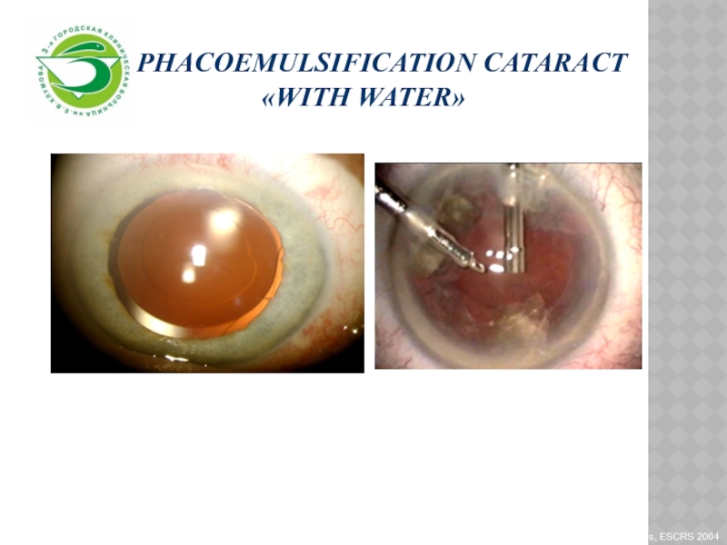
- 16. Phacoemulsification cataract «with water»
- 17. IOL-master for IOL power calculationQuickly and accurately!
- 18. Excellent vision at multiple distances Acrysof
- 19. Toric IOL for precise astigmatism correction mark the cornea prior to implantation…
- 20. New quality of visionToric IOL
- 21. Mark the cornea prior to implantationWith the
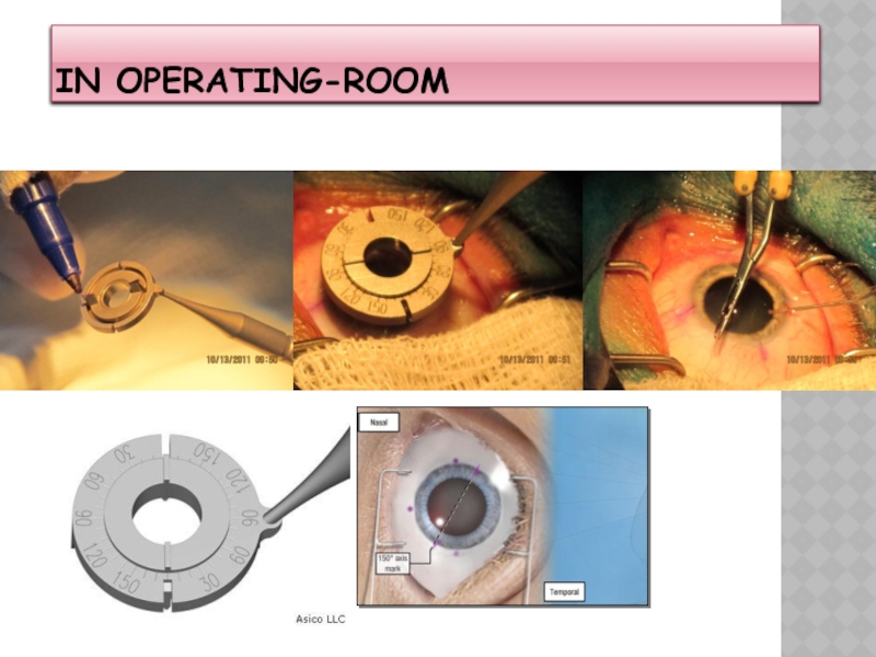
- 22. In operating-room
- 23. Слайд 23
- 24. Implantation of IOL with sulcus fixationIn cases: after eye traumain post-operative aphakiain zonulla thickness
- 25. Vitreo-retinal surgery
- 26. Слайд 26
- 27. AccurusBIOM
- 28. Vitrectomy with endolasercoagulationДо операцииПосле операции
- 29. Before surgeryAfter Surgery
- 30. Corneal transplantationKeratoconusEndothelial-epithelial corneal dystrophy scarring due to keratitis
- 31. Penetrating keratoplastyBefore surgeryAfter surgery
- 32. Скачать презентанцию
Up-to-date diagnostic methods
Слайды и текст этой презентации
Слайд 3Optical Coherence Tomography
Stratus OCT
Non-invasive optical diagnostic techniques that would provide
Слайд 5OCT of anterior eye segment
provides high-resolution anterior chamber images;
measurement of
anterior segment ocular structures,
including anterior chamber depth (ACD), anterior chamber
angles, corneal thicknessallows to evaluate
the potential risk of glaucoma, complications for cataract surgery
Слайд 7The Non-Mydriatic Fundus Camera
Brilliant true color retinal images
Automontage for panoramic
retinal overviews
Documentation of the results and possibility of subsequent management
of the diseaseThe Non-Mydriatic Fundus Camera


