Слайд 1Equine respiratory system diseases
Слайд 2Examination of respiratory system
History taking
Enviromental
Usability of the horse
Enviromental conditio
in stable
Food quality
Dentisity of animals in stable
New animal in stable
Vaccination
Transport
participation
in competitions
History of the disease
How long
Apetite
Animal conditio
Nasal discharge (what type?, how long?, uni/bilateral?)
Cough ( frequency, when)
Dyspnea?
Any treatment?
Слайд 3Examination of respiratory system
General examination
Heart rate, breath rate, lymph nodes,
membrane mucus, temperature
Detail examination of:
Type o breath,
Nasal discharge
Cough
Auscultation of
the larynx, trachea, and chest
Percusion
Aditional tests
Endoscopy (BAL, TW)
USG
X- ray
Endoskopy during exercise
Слайд 4Upper respiratory tract disease
Rhinitis
Necrosis conchae
Polyps
Ethmoid hematoma
Nasal neoplasma
Sinusitis
Pharyngitis
Guttural pouch empyema
Guttural pouch
empyema
Guttural pouch mycosis
Guttural pouch tympany
Guttural pouch chondroids
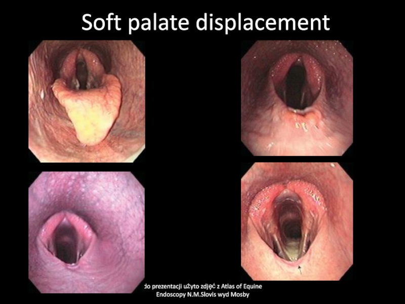
Soft palate displacement
Aryepiglottic fold
displacement
Laryngitis
Laryngeal edema
Larynx neoplasma
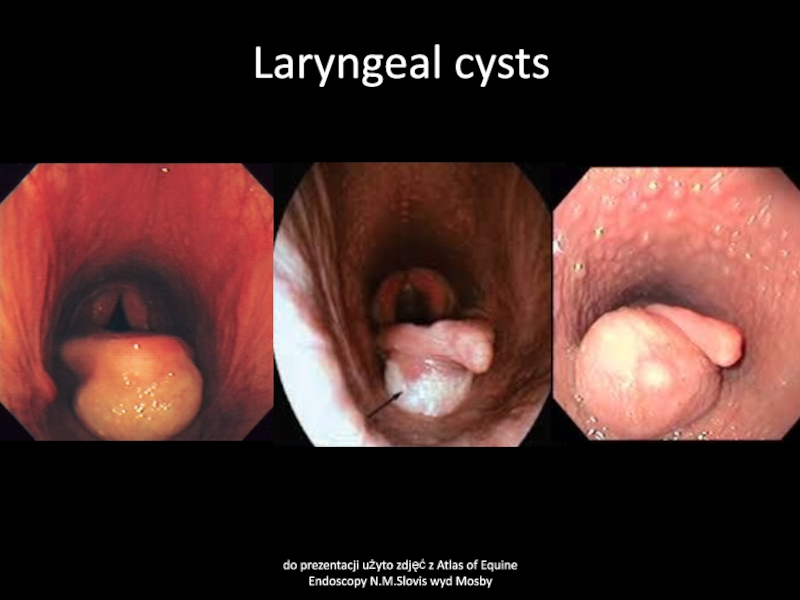
Laryngeal cysts
Laryngeal hemiplegia
Tracheitis & bronchitis
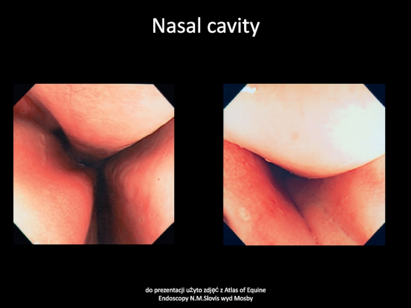
Слайд 5Nasal cavity
do prezentacji użyto zdjęć z Atlas of Equine Endoscopy
N.M.Slovis wyd Mosby
Слайд 6Rhinitis
Cause:
Virus infections-Infuenza, rhinovirus, herpesvirus, arteritis virus, adenovirus, reovirus,
Bacterial infections -Streptococcus
sp., glanders (Psudomonas mallei), other bacteria
Fungi- Aspergillus spp. and many
others different fungi in warm climates
Parasite- Rhinoestrus purpureus, nasal botfly,
Physical factors- dust, smoke, foreign bodies, cold, mechanical trauma (stomach tube, endoscopy) secondary in tumors,
Clinical signs
Nasal discharge (uni/bilateral- serosus, mucosus, purulent, bloody,
Edema,
Pathological respiratory sound, dyspnea, nodules and ulceration (fungus infections)
Decreases performance?
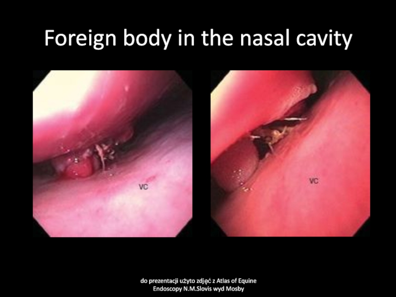
Слайд 7Foreign body in the nasal cavity
do prezentacji użyto zdjęć z
Atlas of Equine Endoscopy N.M.Slovis wyd Mosby
Слайд 8Rhinitis
Clinical pathology
Virology
Bacteriology
Mycology
Mainly to exclude or confirm infectious disease.
In some cases
endoscopy is necessary to find the cause rhinitis
Treatment
Remove primary cause
if possible. Usually self limited illness if primary cause was removed.
Слайд 9Necrosis conche
Cause
Bacterial or fungal infections.
Clinical signs
Muco-purulent, sometimes blood tinged,
odorous discharge uni/bilateral.
Clinical pathology
bacteriology, biopsy, endoskopy
Treatment
removing via endoscopy necrotic
parts of conche. Washing nasal cavities with antimicrobial solutions
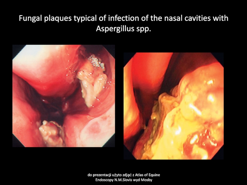
Слайд 10Fungal plaques typical of infection of the nasal cavities with
Aspergillus spp.
do prezentacji użyto zdjęć z Atlas of Equine Endoscopy
N.M.Slovis wyd Mosby
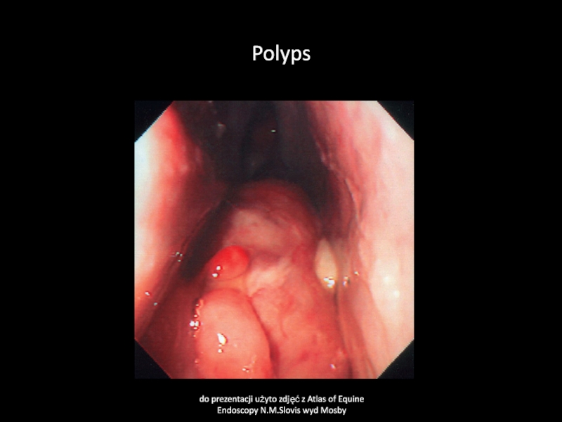
Слайд 11Polyps
Cause
Chronic inflamation of nasal mucous membranes of any cause
Clinical sign
Sero-muco-purulent
nasal discharge uni/bilateral, pathological respiratory sound
Clinical pathology
Biopsy, endscopy
Treatment
surgery
Слайд 12Polyps
do prezentacji użyto zdjęć z Atlas of Equine Endoscopy N.M.Slovis
wyd Mosby
Слайд 13Nasal neoplasma
Cause
Neoplasia- myxoma, fibroma, chondroma, osteochondroma, carcinoma, melanoma
Clinical signs
Uni/bilateral nasal
discharge, sero-muco-purulent, blood tinged, bone deformations, abnormal respiratory sounds, odor,
may be dyspnea
Clinical pathology
Endoscopy, biopsy
Treatment
Surgery, usually poor prognosis
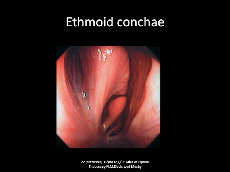
Слайд 14Ethmoid conchae
do prezentacji użyto zdjęć z Atlas of Equine Endoscopy
N.M.Slovis wyd Mosby
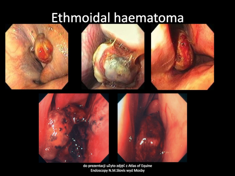
Слайд 15Ethmoidal hematoma
Cause
Neoplasia? Chronic infections, circulatory defect
Clinical signs
At the beginning usually
unilaterally nasal discharge, sero-muco-purulent later blood tinged. Pathological respiratory sounds.
May cause severe dyspnea.
Clinical pathology:
endoscopy, biopsy
Treatment :
surgery, medical treatment-often repeated formalin or alcohol injection intra tumor
Слайд 16Ethmoidal haematoma
do prezentacji użyto zdjęć z Atlas of Equine Endoscopy
N.M.Slovis wyd Mosby
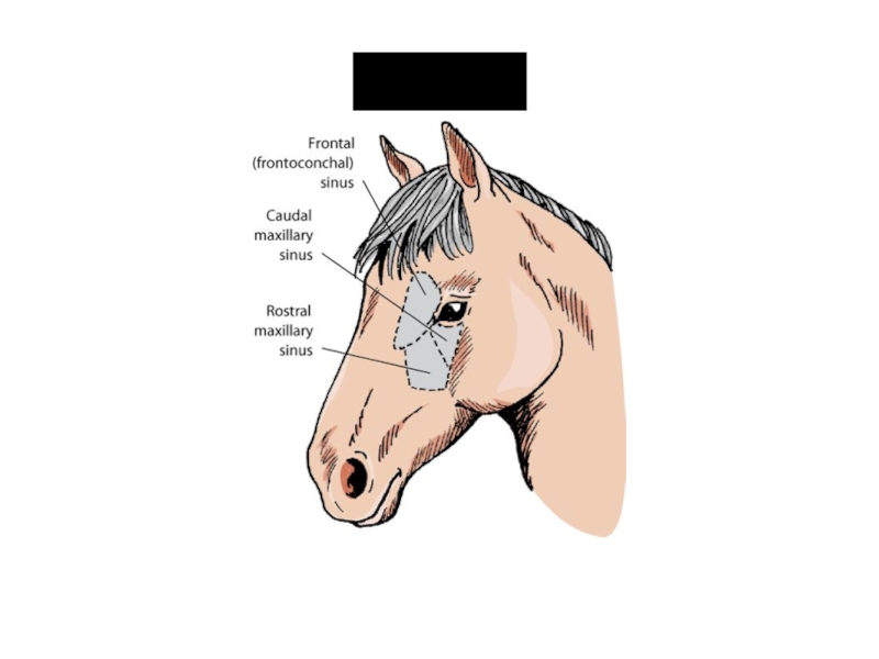
Слайд 18Sinusitis
Cause
Usually secondary to rhinitis, tooth problems, defects of sinus communication
with nasal cavity
Clinical signs:
nasal discharge, uni/bilaterally, more obvious when head
down, sero-muco-purulent, sometimes blood tinged, may be odorous, sinus bone deformity may be visible
Clinical pathology:
bacteriology, mycology, X-ray examination, trepan
Treatment
Surgical opening of sinus, removing the primary cause, antimicrobials, NSAIDs
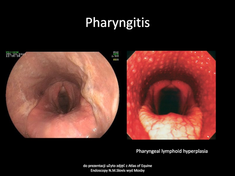
Слайд 21Pharyngitis
Cause
Viral infections- influenza, herpesvirus, adenovirus, arteritis virus,
Bacterial infection-mainly Streptococcus spp.
Physical
trauma-stomach tube, endoscopy, foreign body, chemicals
Слайд 22Pharyngitis
Clinical signs
decreased appetite, difficult swallowing, cough, increased temperature of swollen,
painful throat and local lymhnodes. Nasal discharge- muco-purulent.
Clinical pathology
bacteriology, endoscopy
Treatment
antimicrobials
and NSAIDs
Слайд 23Pharyngitis
do prezentacji użyto zdjęć z Atlas of Equine Endoscopy N.M.Slovis
wyd Mosby
Pharyngeal lymphoid hyperplasia
Слайд 24Pharyngeal paralysis
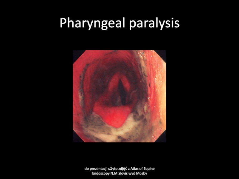
do prezentacji użyto zdjęć z Atlas of Equine Endoscopy
N.M.Slovis wyd Mosby
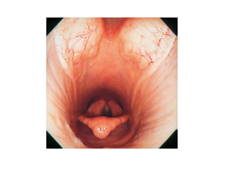
Слайд 25Guttural pouch
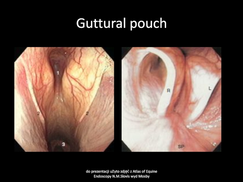
do prezentacji użyto zdjęć z Atlas of Equine Endoscopy
N.M.Slovis wyd Mosby
Слайд 26Guttural pouches
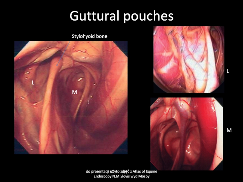
do prezentacji użyto zdjęć z Atlas of Equine Endoscopy
N.M.Slovis wyd Mosby
Stylohyoid bone
M
L
L
M
Слайд 27Guttural pouch mycosis
Cause:
Fungal infections- Aspergillus fumigatus often with bacterial contamination-
Pseudomonas aeruginosa.
Primary lesion in guttural pouch arteries may be the
cause of secondary fungal infection.
Clinical signs:
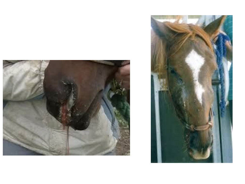
bleeding from nostris, starting from some drops of blood up to severe hemorrhage, usually unilateral. May cause death of animal due to blood loss.
Lesions in nerve in wall of guttural pouch may lead to pharynx dysfunction.
Horner syndrome
Soft palate displacement
Laryngeal hemiplegia
Even fungal encephalitis
Слайд 28Mycosis of the guttural pouches
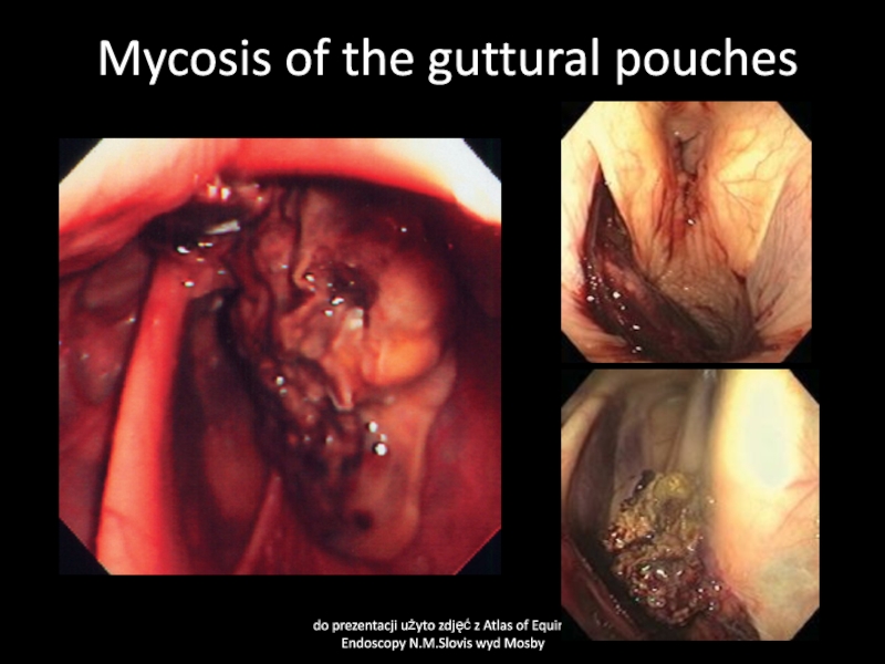
do prezentacji użyto zdjęć z Atlas
of Equine Endoscopy N.M.Slovis wyd Mosby
Слайд 29Guttural pouch mycosis
Clinical pathology
Endoscopy
Mycolgy
Bacteriology
Hematology
Treatment
Local washing with antifungal drugs (econazol, eniconazol,
myconazol, nystatin, natamycin)
Occluding of artery internal or external (branches) by
ballloon or external surgery
Слайд 30Guttural pouch empyema
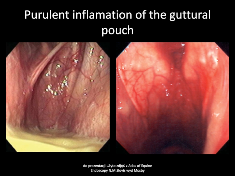
Cause:
mainly Streptococcus spp. Infections,
Clinical signs:
Uni/bilateral muco-purulent nasal discharge,
more obvious when head down. Sweling of guttural pouch region.
Local lymhnodes swollen.
Clinical pathology:
bacteriology, endoscopy
Treatment:
washing out guttural pouch content using normal saline. Antimicrobials
Слайд 31Purulent inflamation of the guttural pouch
do prezentacji użyto zdjęć z
Atlas of Equine Endoscopy N.M.Slovis wyd Mosby
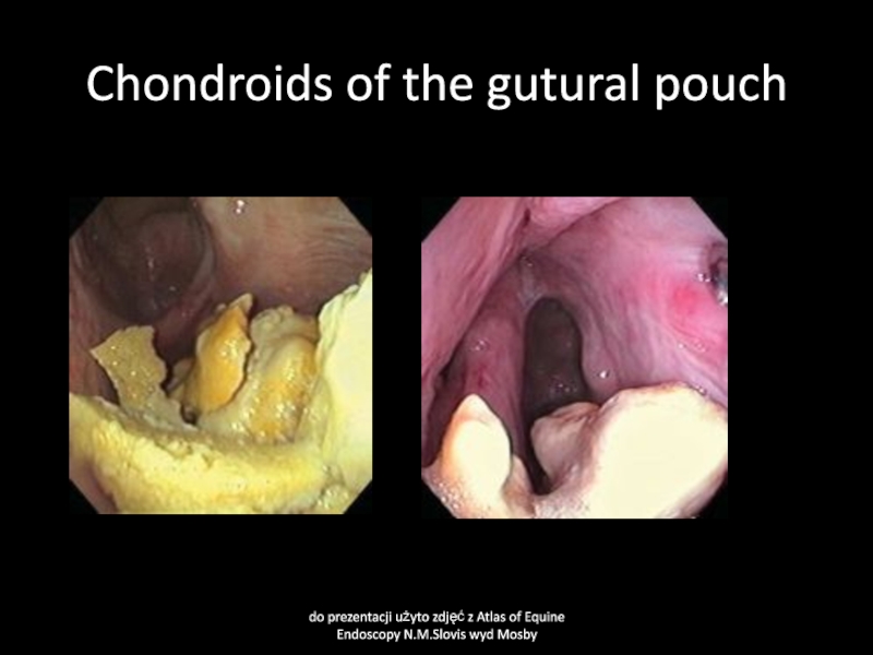
Слайд 32Guttural pouch chondroids
Cause
Inspissated guttural pouch exudate forms stones
Clinical signs
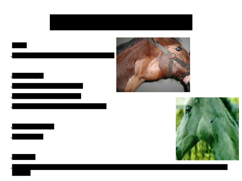
Swelling of
guttural pouch region and typical sound during movement of the
horse head, palpable by hand pressing of guttural pouch
Clinical pathology
Not necessary
Treatment:
Surgery, possible dissolving by acetylcysteine
Слайд 33Chondroids of the gutural pouch
do prezentacji użyto zdjęć z Atlas
of Equine Endoscopy N.M.Slovis wyd Mosby
Слайд 34Guttural pouch tympany
Cause
Congenital defects of guttural pouch operculum
Clinical signs
Swelling of
guttural pouch region
Tympany detected by percusion
May cause difficult swallowing and
dyspnea
Clinical pathology
Not necessary
Treatment
Surgical fistula between pouch in case of unilateral tympany or pharyngeal fistula in case of bilateral tympany
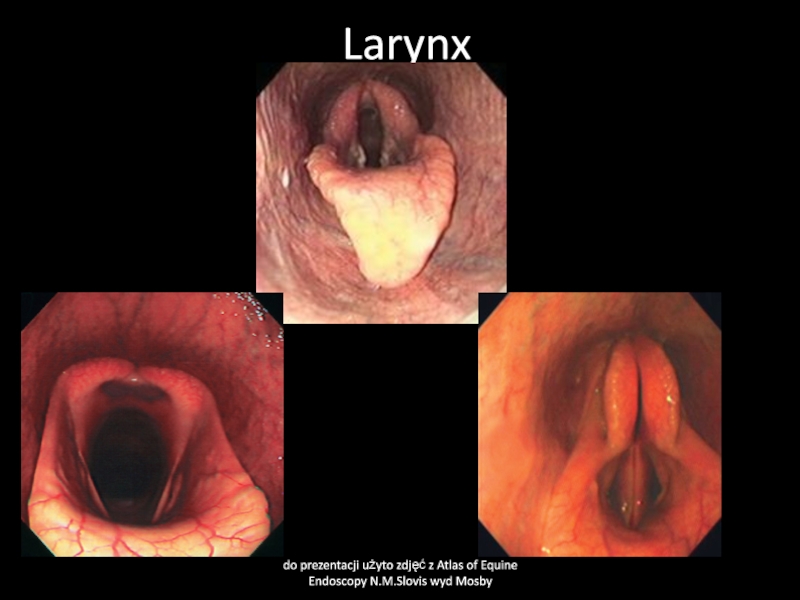
Слайд 35Larynx
do prezentacji użyto zdjęć z Atlas of Equine Endoscopy N.M.Slovis
wyd Mosby
Слайд 36Soft palate displacement
Cause
Paresis of soft palate due to some neurological
deficit, swelling of soft palate, defects of epiglottis and other
umknown reason
Clinical signs: Decreased performance, abnormal respiratory sounds, dyspnea during exercise
Clinical pathology
Endoscopy
Treatment
Anti-inflamatory drugs (flunixin), surgery
Слайд 37Soft palate displacement
do prezentacji użyto zdjęć z Atlas of Equine
Endoscopy N.M.Slovis wyd Mosby
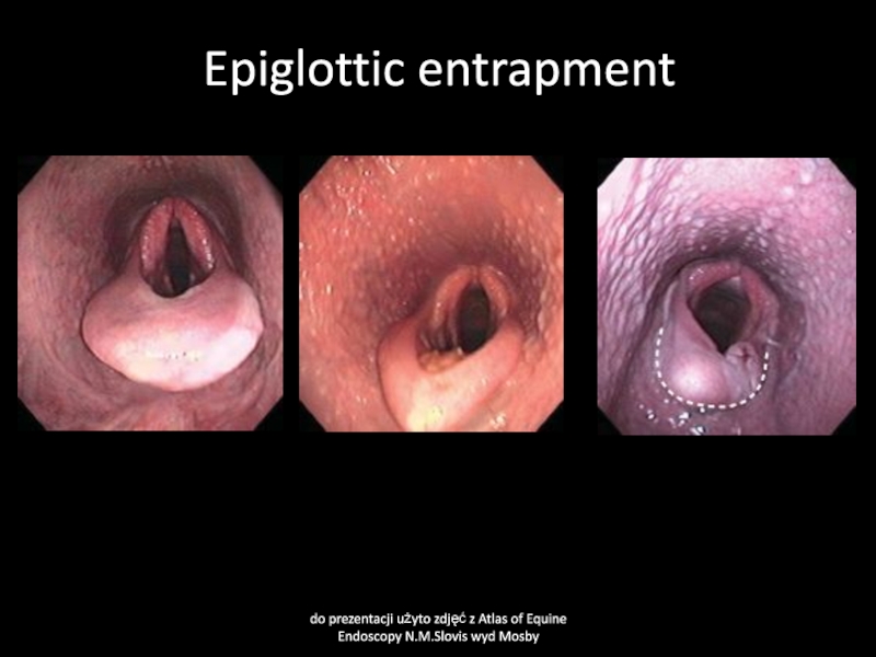
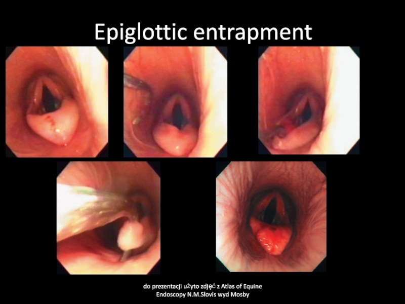
Слайд 38Aryepiglottic fold displacement
(epiglottic entratment)
Cause
Edema of soft tissue close to epiglottis.
Congenital shortening of epiglottis
Clinical signs
Abnormal respiratory sound. Dyspnea during exercise.
Decreased performance.
Clinical pathology
endoscopy
Treatment
surgery. Anti-inflamatory drugs
Слайд 39Epiglottic entrapment
do prezentacji użyto zdjęć z Atlas of Equine Endoscopy
N.M.Slovis wyd Mosby
Слайд 40Epiglottic entrapment
do prezentacji użyto zdjęć z Atlas of Equine Endoscopy
N.M.Slovis wyd Mosby
Слайд 41Laryngitis
Cause
Viral infections- influenza, herpesvirus, adenovirus, arteritis virus,
Bacterial infection-mainly Streptococcus spp.
Physical
trauma-stomach tube, endoscopy, foreign body, chemicals
Clinical signs
Cough, abnormal respiratory sounds,
painful palpation of laryngeal region. Painful swallowing. In some cases fever, decreased appetitte. May cause larynx edema.
Clinical pathology
Endoscopy
Treatment
NSAIDs, antimicrobials in case of bacterial infections
Слайд 42Laryngeal edema
Cause
Acute inflamation, allergy, irritant substances, surgery at larynx region
Clinical
signs
Abnormal respiratory laryngeal sounds, cough, dyspnea, cyanosis, in severe cases
death
Clinical pathology
Endoscopy
Treatment
Steroids, anti-histamine drugs, in some cases tracheotomy. In case of anaphylaxis epinephrine
Слайд 43Larynx neoplasms
Cause
Neoplasia-papilloma, carcinoma, adenoma, fibroma, chondroma
Clinical signs:
Nasal discharge- muco-purulent, blood
tinged, often odorous. Abnormal respiratory sounds, cough, dyspnea, difficult swallowing
Clinical
pathology
Biopsy, endoscopy
Treatment
Surgery, poor prognosis
Слайд 44Larygeal cysts
Cause
Usually congenital cyst
Clinical signs:
abnormal laryngeal respiratory sound, dyspnea, cough
Clinical
pathology
Endoscopy
Treatment
surgery, good prognosis
Слайд 45Laryngeal cysts
do prezentacji użyto zdjęć z Atlas of Equine Endoscopy
N.M.Slovis wyd Mosby
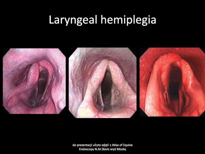
Слайд 46Laryngeal hemiplegia
Cause
Recurrent laryngeal nerve paralysis due to general neuropathy, inherited,
poisonings, local swelling, fungal guttural pouch inflamation
Clinical signs
Abnormal laryngeal respiratory
sounds usually heard only in time of exercise,
Clinical pathology
Endoscopy
Treatment
surgery
Слайд 47Laryngeal hemiplegia
do prezentacji użyto zdjęć z Atlas of Equine Endoscopy
N.M.Slovis wyd Mosby
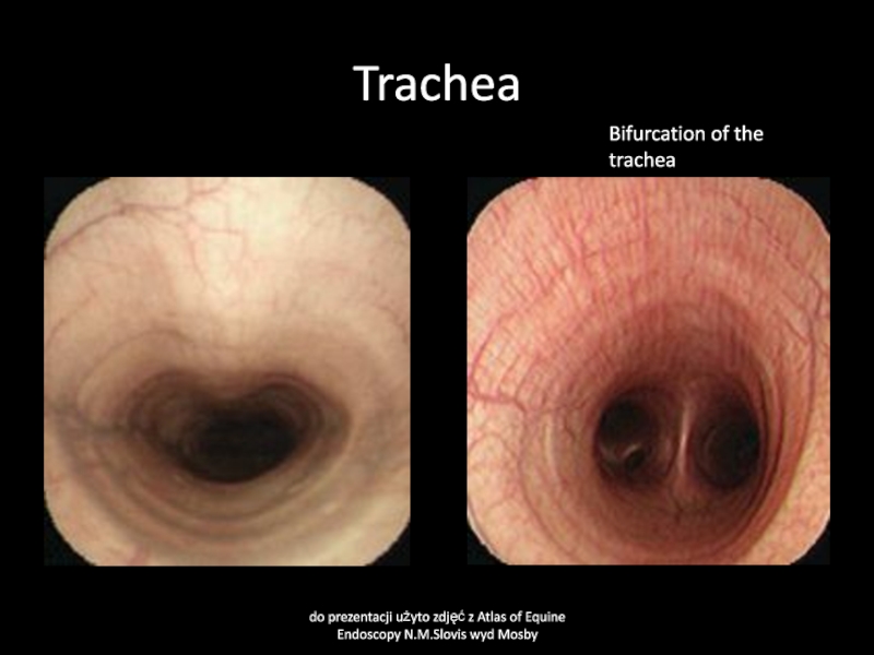
Слайд 48Trachea
do prezentacji użyto zdjęć z Atlas of Equine Endoscopy N.M.Slovis
wyd Mosby
Bifurcation of the trachea
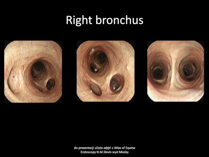
Слайд 49Right bronchus
do prezentacji użyto zdjęć z Atlas of Equine Endoscopy
N.M.Slovis wyd Mosby
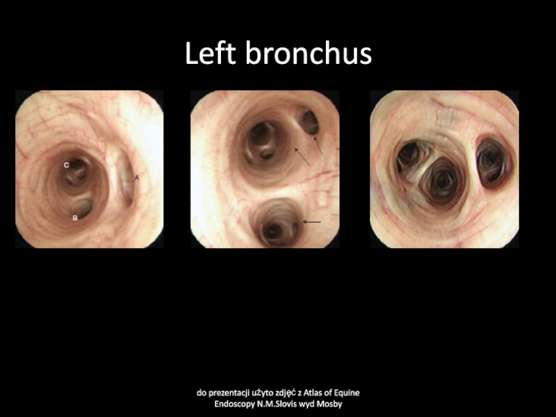
Слайд 50Left bronchus
do prezentacji użyto zdjęć z Atlas of Equine Endoscopy
N.M.Slovis wyd Mosby
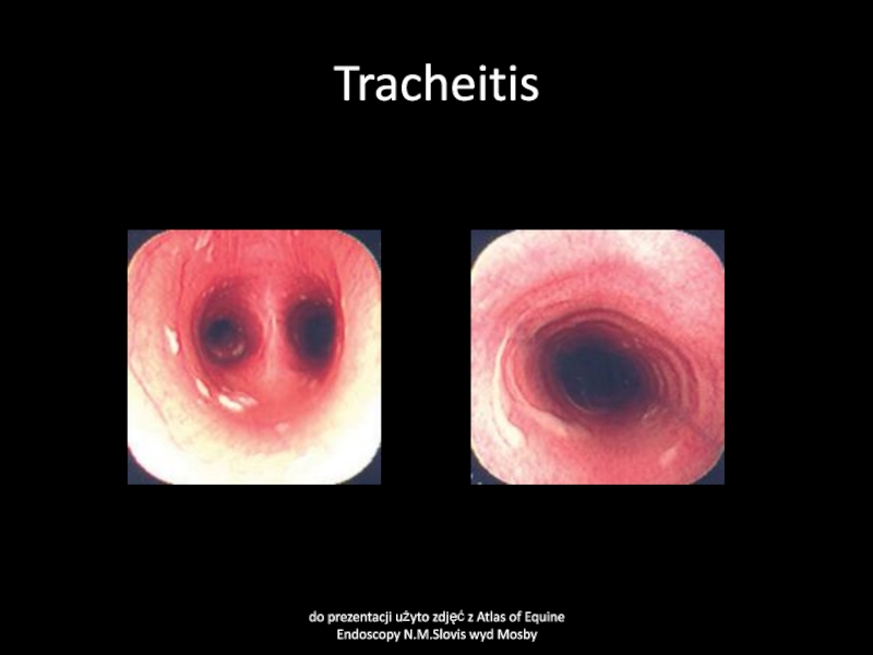
Слайд 51Tracheitis and bronchitis
Cause
Infection equine influenza, equine herpes virus, equine viral
arteritis, streptococcal infections, other bacteria due to stress factors, transport,
contact with new animals, poor hygiene
Clinical signs
May be increased respitration rate, cough, fever, nasal discharge, abnormal respiratory sounds over trachea and lung area, normal result of thorax cavity percusion
Слайд 52Tracheitis
do prezentacji użyto zdjęć z Atlas of Equine Endoscopy N.M.Slovis
wyd Mosby
Слайд 53Exudatives in tracheitis
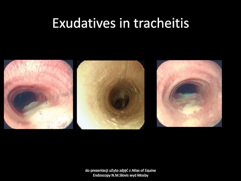
do prezentacji użyto zdjęć z Atlas of Equine
Endoscopy N.M.Slovis wyd Mosby
Слайд 54Tracheitis and bronchitis
Clinical pathology
bacteriological examination of tracheal wash or tharcheal
aspirates, cytology, X-ray, thorax cavity ultrasonography
Treatment
Anitimicrobials
Nsaids
Mucolytics (bromhxine)
rest
Слайд 55Diseases of lungs
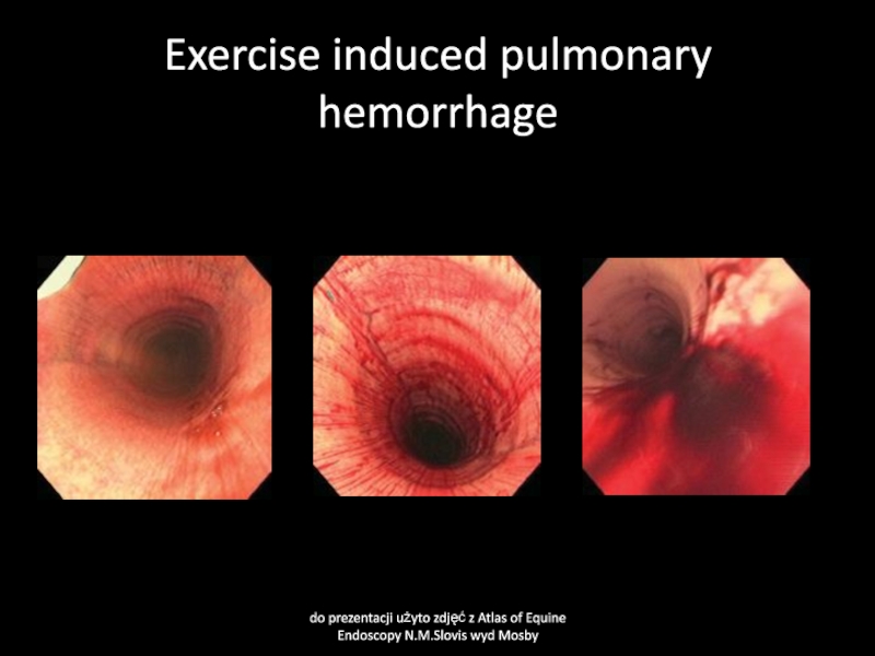
Exercise-induced pulmonary hemorrhage
Recurrect airway obstruction
Слайд 56Exercise-induced pulmonary hemorrhage
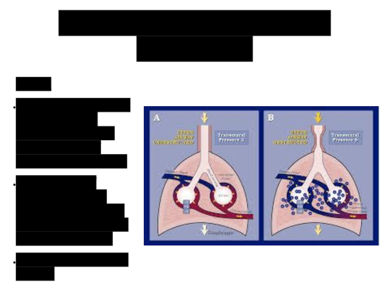
Cause
High pulmonary blood pressure during sternuous exercise cause
rupture of pulmonary capillares.
Possible role of inflamation, small bronchial obstruction
and high intrathoracic negative pressure.
Most common in race horses.
Слайд 57Exercise-induced pulmonary hemorrhage
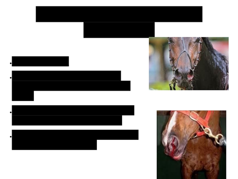
Clinical signs:
May be found in >80% racing horces
but clinically observed in 1-3%.
Sudden slow during race, cough, swallowing
of blood, epistaxis
Some horse may collapse and die due to severe bleeding
Слайд 58Exercise-induced pulmonary hemorrhage
Clinical pathology
Macrophages with digested red blood cells (hemosiderin)
in sample of tracheal aspirates or BAL (broncho-alveolar lavage)
Endoscopy examination
may show blood in trachea or bronchi.
Слайд 59Exercise induced pulmonary hemorrhage
do prezentacji użyto zdjęć z Atlas of
Equine Endoscopy N.M.Slovis wyd Mosby
Слайд 60Exercise- induced pulmonary hemorrhage
Treatment
Rest,
Treat respiratory disease if present.
Furosemide before sternuous
exercise may prevent bleeding, but not allowed in some countries
before the race
Vit K and C
Слайд 61Recurrect Airway Obstruction (Heaves)
Cause
Dusty stable environment, viral infections, air pollution
by Aspergillus fumigatus
Actinomyces spp. And other antigens, allergens at summer
pasture.
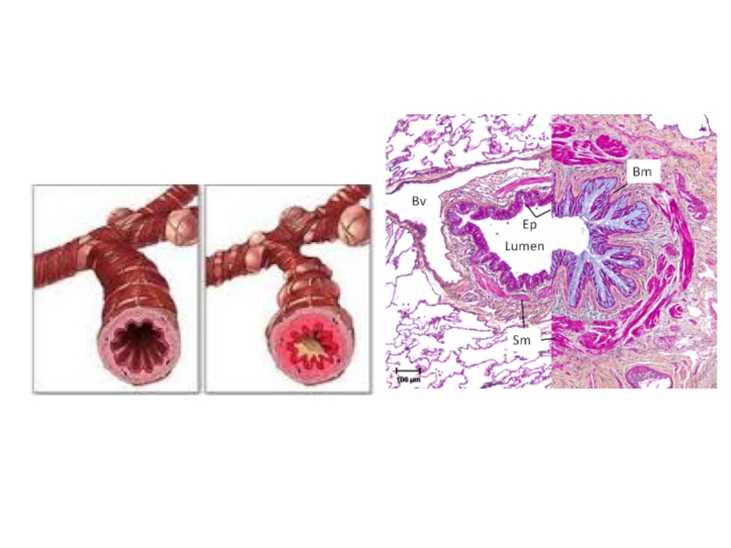
All these factors cause allergic respiratory tract reaction, mainly in small bronchioles
Слайд 64Recurrect Airway Obstruction (Heaves)
Clinical signs:
Older than 7 years horses most
common affected.
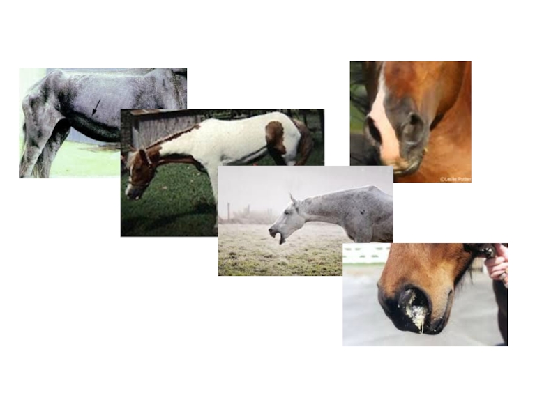
At the beginning cough and nasal discharge which disappeared
after treatment,
Next usually more and more often episode of similar diseases but less curable
After that persistent cough, nasal discharge, increased respiratory rate, expiratory dyspnea („heave line” due to supporting action abdominal muscle during expiration).
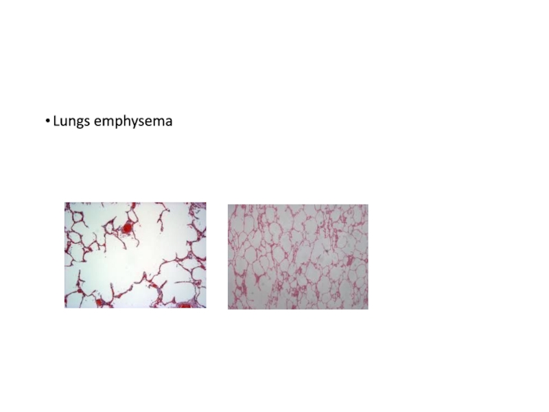
Abdominal sound (wheezing and cracling) on thorax ausculation, abnormal result of thorax percusion (increased resonance at upper caudal part of lung area)
Слайд 66Recurrect Airway Obstruction (Heaves)
Clinical pathology
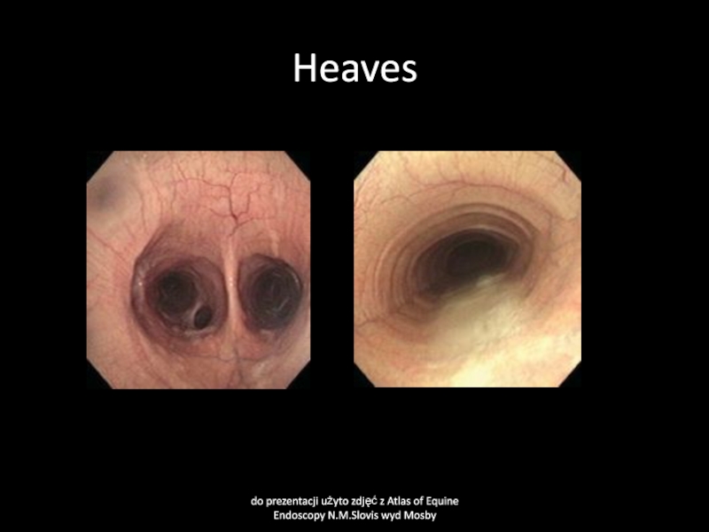
Endoscopy examination ( chronic inflamation of
bronchi and tracheal mucosa visible)
Bronchoalveolar lavage (BAL) contains neutrophils, usually
> 50% and few macrofages
Слайд 67Heaves
do prezentacji użyto zdjęć z Atlas of Equine Endoscopy N.M.Slovis
wyd Mosby
Слайд 68Recurrect Airway Obstruction (Heaves)
Treatment
Change the envionment of the horse,
Wood shavings
instead od straw as a bedding.
Wetted hay
Corticosteroids, bronchodilators (clenbuterol)
orally or as inhalation.
Without change of dusty environment successful treatment is impossible, drugs will only diminish the severity of clinical signs and allow to use the horse a little longer

